Association between Body Condition and Oxidative Status in Dogs ()
1. Introduction
In recent years, Oxidative Stress (OS) has been postulated to be an important factor in the pathogenesis and development of lifestyle-related diseases [1]. In aerobic cells, free radicals are constantly produced mostly as Reactive Oxygen Species (ROS). Once produced, free radicals are removed by antioxidant defences including endogenous and exogenous antioxidants. The imbalance between prooxidant and antioxidant defences in favor of prooxidants results in oxidative stress associated with the oxidative modification of biomolecules such as lipids, proteins, and nucleic acids. Alone or in combination with primary etiological factors, free radicals are involved in a pathogenesis of many inflammatory, degenerative and neoplastic diseases [2,3]. Recently, the prevalence of obesity has been related to a decrease of the plasma antioxidants [4-6], the accumulation of fat has been related to the increase of oxidative stress markers [1,4,6], the oxidative stress has been related to high body mass index (BMI) in human [7] and, finally, obesity has been related to chronic inflammatory status [8]. When caloric intake exceeds energy expenditure, the substrate-induced increase in Krebs cycle activity generates an excess of ROS [8]. Energy imbalances lead to the storage of excess energy in adipocytes, resulting in both hypertrophy and hyperplasia. These processes are associated with abnormalities of adipocyte function, particularly mitochondrial stress and disrupted endoplasmic reticulum function [9]. This adipogenesis implies the differentiation of preadipocytes into mature and secreting adipocytes which release a large number of cytokines, termed “adipokines”. In addition, the enlargement of adipocytes by fat storage induces adipose tissue hypoxia and the secretion of high levels of inflammatory cytokines [8]. The activation of inflammatory signaling pathways in turn increases mitochondrial ROS generation [9]. Although oxidative stress is considered the underlying mechanism by which dysfunctional metabolism occurs in obese subjects, there are still very few studies both in feline and canine subjects [10- 12]. The purpose of this study was to evaluate simultaneously the effects of diet on body weight and on the oxidative and inflammatory status in a group of adult dogs.
2. Materials and Methods
2.1. Animals and Experimental Design
A group of 12 healthy adult German Shepherd dogs, all living together in the same environment, was fed with a maintenance diet (diet A), integrated with natural antioxidants, for a period of 6 months. The amount of feed administered was calculated according to the body weight, in accordance with what is indicated by the manufacturer (13.5 g/Kg bw/day).
The transition to new diet was gradual and occurred in about a week. At the beginning and the end of the trial, every dog was weighed and its BCS was evaluated. In addition, each dog was subjected to a blood sample for the determination of haematological, oxidative and inflammatory parameters. Previously, the same dogs were fed with a different maintenance diet (diet B) for a period of at least 6 months, at the dose of 15 g/Kg bw/day.
2.2. Feed Formulation
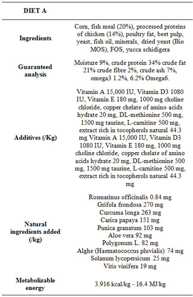
The nutritional values, on the label, of the dry food of the trial (diet A) and of the previous one (diet B), are collected in Tables 1 and 2.
2.3. Evaluation of Body Condition Score (BCS)
The BCS was assessed by a 5-points scale [13].
2.4. Sampling
Blood was sampled from each dog, fasted for 12 hours, after a clinic examination, at the end of each period of six months of diet. CBCs were assessed on EDTA blood sample within 1 hour from the sampling. Plasma and serum samples were obtained after centrifugation at 2400 rpm for 10 min and were stored at −18˚C until further analysis.
2.5. Laboratory Analysis
To evaluate oxidative and inflammatory status some parameters were assessed: CBCs (Complete Blood Counts), d-ROMs (derivatives of Reactive Oxygen Metabolites), BAP (Biological Antioxidant Potential), Retinol, α-to-copherol, Fibrinogen and CRP (C Reactive Protein).
CBCs was determined by IDEXX Lasercyte® hematology analyser (IDEXX Laboratories, Inc., Westbrook, Maine, USA) that provides complete blood counts including a true five-part white blood cell differential and absolute reticulocyte count. Morphological evaluations of blood cell populations were made through the observation of a peripheral blood smear.
Wo

Table 1. Nutritional values of maintainance diet administerred to dogs for 6 months.

Table 2. Nutritional values of diet consumed by dogs before the trial for at least 6 months.
d-ROMs and BAP plasma concentrations were determined by a spectrophotometric methods (Slim, SEAC, Florence, Italy). In the d-ROMs test (Diacron International, Grosseto, Italy), the reactive oxygen metabolites (hydroperoxides primarily) of a biological sample, in the presence of iron released from plasma proteins by an acidic buffer, are able to generate alkoxyl and peroxyl radicals according to the Fenton’s reaction. Such radicals, in turn, are able to oxidize an alkyl-substituted aromatic amine (N,N-dietylparaphenylendiamine), thus producing a pink-coloured derivative which is photometrically quantified at 505 nm. The d-ROMs concentration runs directly parallel with colour intensity and is expressed as Carratelli Units (1 CARR U = 0.08 mg hydrogen peroxide/ dL). In the BAP test (Diacron International, Grosseto, Italy), the addition of a plasma sample to a coloured solution (thiocyanate) that is obtained by mixing a ferric chloride solution with a thiocyanate derivative solution, causes a colour change. The intensity of the decolouration is measured photometrically at 505 nm and is proportional to the ability of plasma to reduce ferric ions. The results are expressed as μmoli/L. The normal reference range in dogs is 56.4 - 91.4 U.CARR and 1440 - 3260 μmoli/L for d-ROMs and BAP, respectively [14].
α-tocopherol and retinol serum concentrations were analysed by HPLC and UV detection using a commercial test kit (Chromsystems Instrument and Chemical Ltd., Munchen, Germany). The HPLC system consisted of a Series 200 Perkin Elmer gradient Pump (Norwalk, CT, USA) coupled to a Series 200 Perkin Elmer variable UV detector (Norwalk, CT, USA), which was set at 325 nm and 295 nm. HPLC was coupled to a personal computer by using an interface SERIES 600 Perkin Elmer (Norwalk, CT, USA). Integration of peaks was performed through Turbochrome Navigator software (Perkin Elmer, Norwalk, CT, USA). The results were expressed as μg/mL.
Fibrinogen was determined by coagulometric method (Clot2, SEAC, Calenzano, Italy). The quantitative determination of fibrinogen is based on the addition of a relatively large amount of thrombin to diluted citrate plasma, so that the clotting time depends only on the fibrinogen contained in the sample. The assay procedure consists of placing 200 μL of 1:10-diluted plasma in a test tube preheated to 37˚C, followed by incubation for 2 minutes at 37˚C, and then adding 200 μL of the fibrinogen reagent (preheated to 37˚C). Upon the addition of the fibrinogen reagent, the stopwatch was started, and the clotting time was measured. For this assay, the results in seconds must be converted into mg/dL using a conversion table supplied with the kit.
CRP was assessed by immunoturbidimetric test (Beckman Coulter s.r.l., Milan, Italy). CRP is an acute phase protein synthesized by the liver in response to the release of inflammatory cytokines such as interleukin-6. The concentration of CRP increases significantly as a result of acute or chronic inflammation observed in the case of bacterial infections (the most powerful stimulus for the CRP production), an autoimmune disease or immune complex, tissue necrosis and malignant tumors, myocardial infarction and trauma. The increase is recorded by 24 to 48 hours and the level can be 2000 times higher than the normal value. In many cases, the changes in plasmatic CRP level are preceding the clinical symptoms. When a sample is mixed with the buffer solution and antiserum solution, the CRP reacts specifically with anti-CRP giving rise to insoluble aggregates. The absorbance of these aggregates is proportional to the concentration of CRP in the sample.
2.6. Statistical Analysis
The data were examined for normality on the basis of Kurtosis and Skewness coefficients. t Student test was applied to evidence differences in the investigated parameters, between before and after the trial; Pearson multiple correlation test was applied to measure the strength of the linear relationship between the variables. Results were considered significant at a value of p < 0.05. Statistical analysis was performed by use of commercial statistical software (STATGRAPHICS Plus®, Centurion).
3. Results
All the data were normally distributed, except CRP to which has been applied the transformation log10. Table 3 contains data before and after the administration of diet A. At the beginning of the period, 83% of dogs showed a BCS greater than 3 while 17% was attributed a BCS

Table 3. Data (M ± SD) of analysed parameters before and after the administration of diet A.
slightly less than 3. At the end of the trial, all of the subjects reached the ideal weight (p < 0.01). All haematological parameters were found to be within the reference range in both controls; in Table 3 have been reported those which showed a significant variation to vary the diet (HCT and PLT). High significant differences have emerged for d-ROMs, Retinol and PLT; significant differences for BCS, BAP and HCT; no differences for α- tocopherol, fibrinogen and CRP.
Table 4 report Pearson coefficient values emerged by multiple analysis. A weak negative correlation has emerged between BAP vs d-ROMs and CRP vs HCT; a weak positive correlation between number of lymphocytes vs Retinol; a moderate negative correlation between BCS vs BAP, BAP vs α-tocopherol, retinol vs α-tocopherol and HCT vs BAP; a moderate positive correlation between BCS vs α-tocopherol, BAP vs Retinol and BAP vs number of lymphocytes.