Bilateral Central Serous Chorioretinopathy (CRSC) in Pregnancy Complicated with Retroplacental Hematoma: About a Case ()
1. Introduction
Central serous chorioretinopathy (CSCR) first described in 1866 by the German Albrecht Von Graefe under the name of recurrent central retinitis [1] , is a condition characterized by serous sensorineural retinal detachment in the macular region [2] , which is thought to be due to focal leakage at the level of the retinal pigment epithelium [3] . It preferentially affects men than women with an order ratio of 6/1 [4] . The average age of patients with CRSC is 30 - 40 years for men while for women it varies around 40 - 50 years [5] . Certain predisposing factors have been mentioned by the authors, namely: corticosteroids, the frequency of which is approximately 20% in classic CRSCs [6] and 33% - 44% in cases associated with corticosteroid therapy [7] [8] .
The contributing factors mentioned are: corticosteroids, A personalities, arterial hypertension, pregnant women whose risk during pregnancy may be due to the high presence of cortisol levels in the blood. Some studies implicate hemodynamic disturbances; and it is during the third trimester that the risk of CRSC is the greatest [6] [9] [10] [11] [12] [13] .
Some pregnancy complications such as retroplacental hematoma, pre-eclampsia associated with CRSC are rarely mentioned in the literature.
We report and describe the clinical diagnosis of a case of brutal bilateral CRSC associated with a retroplacental hematoma in a secondary ophthalmology center with a limited diagnostic and therapeutic technical platform.
2. Observation
It was a 33-year-old pregnant woman; (4 pregnancies, 4 parities, 2 deceased and 1 stillborn)
G4P4V1D2MN1, having developed a retroplacental hematoma on the ground of toxaemia of pregnancy added to 8 months of pregnancy or approximately 34 weeks of amenorrhea of evolution. She had a sharp drop in visual acuity on admission, blood pressure was 120/80mm Hg, proteinuria was a cross. She was Caesarized on the same day with a fresh stillborn baby, she was sent to us for persistent visual acuity loss two days after Caesarean section.
On external examination, there was edema of the face and lower limbs.
Distance visual acuity was, right eye (OD): counts fingers at 1 meter, left eye (LO): counts fingers at 2 meters. On biomicroscopy, there was chemosis in the anterior segment and the intraocular pressure was: 14 mm hg in OD and 24 mm hg in the left eye. At the fundus, there was a significant lifting of the macular region, round, limited wide, approximately 3 papillary diameters and retinal exudates.
Faced with this symptomatology, we evoked the central serous chorioretinopathy associated with a retroplacental hematoma and added toxemia of pregnancy. The following assessments were requested to support our diagnosis, namely: fluorescein angiography, optical coherence tomography, B-mode ultrasound, photo retina. These assessments could not be carried out in real time, due to the lack or inadequacy of this equipment and also due to the general condition of the patient who had limited mobility. The retina photo taken with the MII device Ret Cam (Made In India, Retinoscope Camera) (Photo 1) coupled with the smartphone gave an image of a bubble of significant serous detachment of the macular region on the right and on the left (image a little blurred) (Figure 1 and Figure 2), confirmed the fundus result. Faced with the hemodynamic disorders, the patient was taken care of by the resuscitation team and the obstetricians. On the ophthalmological level, clinical monitoring, without medical or physical treatment, was instituted.
At one month of evolution, the visual acuity was 7/10 in the right eye as in the left eye. The control retinal photo taken during the period revealed a slight macular oedema (Figure 3) and the OCT, performed, showed an almost normal appearance with disappearance of the serous retinal detachment (Figure 4).
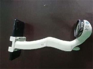
Photo 1. MII Ret Cam device coupled to the smartphone and the volk 20 diopter lens.
![]()
Figure 1. Bulla of macular serous detachment in the right eye (arrow).
![]()
Figure 2. Bulla of serou macular detachment in the left eye (arrow).
![]()
Figure 3. Retinography: Retinal exudates, macular edema after one month.
![]()
Figure 4. OCT after one month of evolution (normal).
Two months later, the control revealed: bilateral visual acuity at 10/10, a slight macular reorganization and some retinal exudates. We will continue this monitoring.
Despite this favorable evolution, monitoring is still the rule, in order to prevent recurrences and complications that could compromise the visual prognosis.
We know that optical coherence tomography (OCT) has, in recent years, revolutionized the diagnostic and therapeutic approach of CRSC. It is an objective, non-invasive and above all quantitative examination, allowing an early and precise diagnosis of CRSC. Thanks to the OCT, a complete assessment of the various retinal and even choroidal lesions present during the CRSC, is now possible with a quantitative follow-up of the serous retinal detachment [14] . But this examination is not always available at the level of secondary health structures in developing countries like ours.
This case testifies that the clinical diagnosis of central serous chorioretinopathy remains possible in the absence of means of explorations such as OCT, angiography.
In the absence of these examinations, the diagnosis is made in the presence of a bubble rising from the macular region to the fundus and the spontaneously favorable evolution without treatment under associated clinical supervision. The association of images from a simple retina photo device such as the MII ret Cam, may be a valuable aid in diagnosis.
3. Discussion
Currently the exact origin of chorioretinopathy is still uncertain, several hypotheses are put forward, according to Gass [6] , the CRSC would be due to a very localized alteration of the pigment epithelium, this would allow the liquid located at the choroidal level to pass under the retina. Spitznas [15] , reports that when CRSC appears, fluid flow changes from a retinochoroidal direction to a choroidoretinal direction. This phenomenon would be due to an inhibition or a reversal of the flow of ions, which will lead to a call for liquid at this location [2] [16] .
Several risk factors related to CRSC have been described in the literature. A 2016 meta-analysis by BING LIU et al. [17] , indicated that hypertension, Helicobacter pylori infection, use of steroids, sleep disorders, autoimmune diseases, use of psychopharmacological drugs, gastroesophageal reflux disease, peptic ulcer. the use of antihistamines, antacids/anti-reflux agents, psychological stress, pregnancy and alcohol consumption were important risk factors linked to the occurrence of CRSC. The authors believe that the level of urbanization could be a potential protective factor for CRSC. However, for some factors further studies would be needed to be confirmed [17] .
For stress, it was reported by F. Chraibi [18] in a case-control study in Morocco in 2017. Previously, Rouvass et al. [19] in the years 2010 to 2011 in Europe, reported an increase in the incidence of new and recurrent cases of CRSC coinciding with the financial crisis. During the Ivorian politico-military crises between 2003 and 2005, Fanny et al. [3] , reported 6 cases of CRSC in a center where no case of CRSC had been diagnosed before the crisis. In our case, was our patient subjected to phenomena of pregnancy stress? Although this aspect has not been taken into account, some studies assess its impact during pregnancy on the mother, the unborn child and on the obstetric outcome [20] . As for Pregnancy, it represents 5.1% to 10% of cases of CRSC among women with [9] [10] , and it is during the 3rd trimester that the risk of CRSC is most frequent [6] [13] . This corroborates with our observation which was at 8 months (3rd trimester) of active pregnancy. This same gestational age period was reported by Chandana C et al. in Calcutta India [21] ; as well as Olousanya BA et al. in Nigeria in whom the gestational age was 29 week amenorrhea [22] . Yu et al.; in a study of the clinical characteristics of pregnancy-associated CRSC in the Chinese population brought relatively the same gestational period (27 ± 2.09 week amenorrhea) from 2012 to 2019 [23] . Despite the marked male predisposition in the incidence of CRSC [17] , pregnancy is a known risk factor and the occurrence of CRSC during pregnancy may be the result of increased endogenous corticosteroid levels associated with pregnancy [24] .
Although arterial hypertension is a risk factor for CRSC, the authors agree that serous retinal detachment is a rare complication of preeclampsia/eclampsia affecting 1% - 2% of preeclamptics [25] .
Our observation had superimposed preeclampsia complicated by retroplacental hematoma; the occurrence of CRSC during a pregnancy complicated by preeclampsia and retroplacental hematoma is rarely reported by the authors. Sibel I et al. [25] reported a case of bilateral serous retinal detachment involving the center of the macula accompanied by intraretinal fluid in a pre-eclamptic pregnant woman, but which was associated with posterior reversible encephalopathy syndrome.
Ngwanou A et al. [26] in Cameroon in a study on retinal lesions during preeclampsia/eclampsia reported that papilledema was the most common retinal lesion in preeclampsia/eclampsia, CRSC was not found in any of the patients.
Our observation had no papilledema, so the pre-eclampsia was a superimposed toxaemia of pregnancy (one-cross proteinuria).
The retroplacental hematoma results from the rupture of the tissue walls following the lifting of a spasm, and creates tissue damage at the level of the basal plate of the uterus [27] . The mechanisms of its involvement in the occurrence of CRSC are not yet described in the literature to our knowledge. This entity could be among the first cases reported and could be explained by the coexistence and severity of risk factors in this patient. Further research may support the role of placental hematoma in the onset or progression of CRSC.
CRSC was bilateral in our patient with a severe loss of visual acuity: counting fingers at 1 meter in the right eye and 2 meters in the left eye. Sibel I and all and Chandana C and all in India also found bilateral involvement in observations with respectively corrected visual acuities of 20/100 on the right, 20/50 on the left and uncorrected visual acuities of 6/60 on the eye right and count fingers 4 yards to left eye; on the other hand, the attack was unilateral with a visual acuity of 6/9 in the left eye in a pregnant woman without associated complications, according to Olusanya BA in Nigeria.
The significant decrease in bilateral visual acuity in our patient could be explained by the severity of the complications incumbent on the retroplacental hematoma which leads to hemodynamic disorders favoring the occurrence of CRSC.
At two months of progression without treatment, bilateral visual acuity was 10/10. This confirms the data in the literature where the visual prognosis during CRSC remains good in the majority of cases in the absence of treatment [28] [29] . Indeed, in the majority of cases there is a complete spontaneous resorption of the serous retinal detachment [30] .
Although most of them heal spontaneously with better visual acuity and improvement of symptoms in 3 months [31] , the recurrence rate can reach 50% without any treatment [32] and 10% of patients with SCC have more than three recurrences when follow-up lasts 15 years [33] , which demonstrate a poor prognosis such as retinal dysfunction and permanent visual loss [34] . Monitoring is still necessary despite this spontaneous recovery.
4. Conclusion
Central serous chorioretinopathy induced by pregnancy is not unusual; however, its bilateral and sudden form in a context of pregnancy complicated by pre-eclampsia and retroplacental hematoma is rare. An ophthalmological consultation should be routinely requested for any 3rd trimester pregnancy with no complications. In the absence of means of exploration such as OCT, angiography, the clinical diagnosis of central serous chorioretinopathy remains possible and is made in front of a bubble of lifting of the macular region at the fundus associating with the images of a simple photo device retina like the MII ret Cam. The evolution is spontaneously favorable under surveillance without other associated treatments.