1. Introduction
Oxygen free radicals or, more generally, reactive oxygen species (ROS) are believed to be involved in the etiology of certain diseases [1] [2] . The biological effects of ROS are controlled in vivo by enzymatic and non-enzymatic mechanisms. But, in some conditions, these effects cannot be controlled and lead cell damages. In such cases, exogenous antioxidants can be useful. Among the drugs, Isoniazid (INH) induces oxidative stress, which is one of the mechanisms responsible for cytotoxicity [3] . For these purposes, natural plant products have been used since ancient times. Salvia officinalis L. (Lamiaceae) is among the plants to which hypolipidaemic properties have been attributed by traditional medicine; it is a common aromatic plant, which is distributed widely in the Mediterranean and the Middle East countries. Studies on the leaves, roots and water soluble extractions of Salvia officinalis L. showed that they contain volatile fatty acids, diterpens, saponins, flavonoids, phenolic acids, resin and oestrogenic substances [4] -[7] . In the present study, we evaluate antioxidant effects of Salvia officinalis L. hydroalcoholic extract and its therapeutic effect to counteract liver damage in male rats.
2. Materials and Methods
Salvia officinalis L. plants were collected from Kordan area of Karaj in Iran. The aerial parts of plants were lyophilized and kept at −20˚C. Hydroalcoholic extract was prepared using food grade ethanol and deionised water (96 and 4%, respectively). Male Wistar rats (180 ± 20 g) were purchased from Tehran Pasteur Institute (Iran) and acclimated to our laboratory animal facilities for at least one week before the start of the experiments. During this period, the animals were maintained on a natural light/dark cycle at 20˚C ± 2˚C and given food and tap water ad libitum. The animals were kept and handled in accordance to our University regulations that follows the Guidelines for the Human Use and Care of Laboratory Animals. The rats were randomly divided into 8 groups (7 rats each) as follows: Group 1, healthy control rats received distilled water as sole drinking source; Group 2 received Isoniazid (INH), 50 mg/kg orally by gavage; Groups 3, 4 and 5 were treated intraperitoneally (IP) with Salvia officinalis L. hydroalcoholic extract with the doses 100, 250 and 400 mg/kg, respectively; Groups 6, 7 and 8 were coadministered with Isoniazid (50 mg/kg) and Salvia officinalis L. hydroalcoholic extract 100, 250 and 400 mg/kg, respectively. All the treatments were done for 28 days. Hepatotoxicity of Isoniazid, and Salvia officinalis L. hydroalcoholic extract effects were then evaluated. Also changes in aspartate aminotransferase (AST), alanine aminotransferase (ALT), alkaline phosphates (ALP), gamma-glutamyltransferase (GGT) levels were assessed. At the end of the experiment, animals were sacrificed by cervical dislocation and blood plasma was collected. A fresh piece of the liver from each rat with approximately 2 mm thickness, was rapidly immersed in Bouin’s solution and kept for 24 h at 4˚C. Fixed tissues were then processed routinely for embedding in paraffin, sectioned (5 µm), deparaffinized and rehydrated using standard techniques. The extent of liver damage was evaluated based on morphological changes in liver sections stained with hematoxylin and eosin (H & E) using standard techniques.
Statistical Analysis
Data are expressed as means ± SEM. The comparison between the means of treatments and control group was performed using Student’s t-test. P values ≤ 0.05 were considered statistically significant.
3. Results
3.1. Effects on Liver Enzymes
Serum level of AST in rats coadministered with both INH and plant extract at 250 and 400 mg/kg showed significant reduction when compared to INH group, that may indicate the antioxidant effects of the plant extract while no differences were seen with control (Figure 1). At all plant extract concentrations, specially at 250 mg/kg, serum ALT level significantly reduced when coadministered with INH compared to both INH and control group (Figure 2). Evaluation of GGT in all groups revealed that the plant extract at doses of 100 and 250 mg/kg alone and with INH 50 mg/kg, has alleviated the enzyme comparing to INH receiving rats (Figure 3). Coadministration of the plant extract at concentrations of 250 and 400 mg/kg with INH again showed significant reduction in ALP serum level statistically (Figure 4).
3.2. Liver Histopathology Study
Histopathology effects of the plant extract on rat liver have been also studied in all groups. All microscopic ob-

Figure 1. Serum level of AST in rats in different groups; s = plant extract, INH = Isoniazid. *: compared to control, +: compared to INH group. P < 0.001.

Figure 2. Serum level of ALT in rats in different groups; s = plant extract, INH = Isoniazid. *: compared to control, +: compared to INH group. P < 0.001.

Figure 3. Serum level of GGT in rats in different groups; s = plant extract, INH = Isoniazid. *: compared to control, +: compared to INH group. P < 0.001.
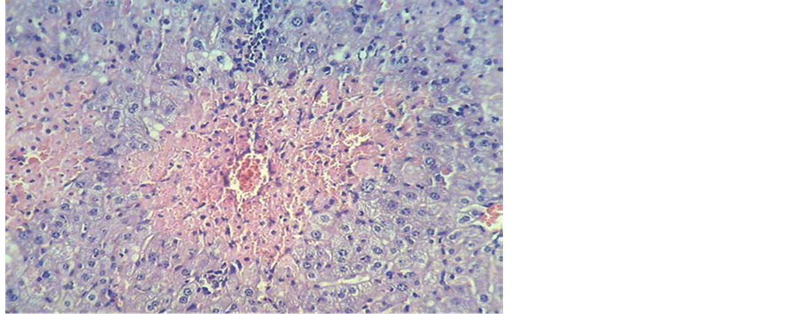
servations have been performed and reported by a pathology expert. Figure 5 shows the normal liver section stained with H & E. After treatment of rats with INH 50 mg/kg for 28 days, severe tissue necrosis, and inflammation of central vein in liver and lymphocyte proliferations were observed (Figure 6). Figure 7 shows the mild dilution in central vein and sinusoids in rat liver received the plant extract 250 mg/kg. Finally the liver histopathology alterations in rats coadministered with the plant extract 250 mg/kg and INH is shown in Figure 8 that indicating low sinusoids dilution.
4. Discussion
In the present study, the protective role of the plant extract on liver has been emphasized. Interestingly, at different plant concentrations, the biochemical parameter changes and histopathology studies revealed marked reduction in INH-induced hepatic damage. In this research, first hepatotoxicities in rats were produced by administering INH (50 mg/kg) orally for 28 days. A significant rise above the normal upper limits in the measured serum transaminases was considered a biochemical indication of liver injury. Increased oxidative stress play a role in the development of liver injury induced by INH [8] [9] . Previous reports in rats suggest that the hydra zine metabolite of INH which has subsequent effect on CYP2E1 induction is involved in the development of

Figure 4. Serum level of ALK in rats in different groups; s = plant extract, INH = Isoniazid. *: compared to control, +: compared to INH group. P < 0.001.

Figure 5. Microscopic photo of normal rat liver stained with H & E ×400.

Figure 6. Microscopic photo of rat liver treated with INH 50 mg/kg stained with H & E ×200.

Figure 7. Microscopic photo of rat liver treated with plant extract 250 mg/kg stained with H & E ×200.

Figure 8. Microscopic photo of rat liver treated with both INH 50 mg/kg and plant extract 250 mg/kg stained with H & E ×200.
INH-induced hepatotoxicity, and also oxidative stress as one of the mechanism for INH induced hepatic injury [8] -[10] .
Oxidative stress is involved in the pathogenesis of many complications in animals and humans. Excessive oxidative stress causes exceeding free radicals product especially reactive oxygen species (ROS). ROS has been known to produce cellular and tissue injury through covalent binding, DNA strand breaking, lipid peroxidation (LPO) and augment fibrosis [11] [12] .
The liver is regarded as one of the central metabolic organs, regulating and maintaining homeostasis. The increase in the level of AST, ALT, ALP and GGT are important indices for hepatic damage. Amino transferases are present in high concentration in liver, an important class of enzymes linking carbohydrate and amino acid metabolism. In cases of liver damage with hepatocellular lesions and parenchymal cell necrosis, these marker enzymes are released from the damaged tissues into the blood stream [10] [12] .
In the present study, the activities of these enzymes were found to increase in the hepatotoxic animals, and were significantly reduced in groups of the plant extract administered rats as compared to that of toxicant rats. This confirms the protective effect of the extract against INH induced hepatic damage. The effect was more pronounced with 250 mg/kg extract.
So far, hepatoprptective effects of many plant extract have been reported. Possible mechanism of all of the extracts including presenting herb as hepatoprotective may be due to their anti-oxidant effects or inhibition of cytochrome P450. This might be due to the high contents of polyphenoles present in the extract which could have reduced the production and/or accumulation of toxic derived metabolites [11] [13] -[19] . Histopathological examination of the liver section of the rats treated with INH showed intense centrilobular necrosis and vacuolization. The rats treated with the plant extract and extracts along with toxicant showed sign of protection against these toxicants to considerable extent as evident from formation of normal hepatic cards and absence of necrosis. The hepatoprotective effect of the extract may be explained depending on the fact that it contains polyphenolic compounds like toyon, cineole and camphor which may scavenge free radicals offering hepatic protection and its antioxidant potential mechanism suggesting that the extract of plant may be useful to prevent the oxidative stress induced damage. More research is required in this point of view to develop a suitable hepatoprotective drug from the plant extract. Purification of extracts and identification of the active ingredient may yield to introduce active hepatoprotective agents.
5. Conclusion
The results of this study indicate that Salvia officinalis L. extract has hepatoprotective action against INH induced hepatic damage in rats. This study suggests that possible mechanism of this activity may be due to free radical-scavenging and antioxidant activities which might be attributed to the presence of flavonoids in the extract.
NOTES
*Corresponding author.