
1. Introduction
Successful prosthetic rehabilitation requires an understanding of the biomechanics involved in establishing and maintaining prosthetic stability, support and retention (Housset’s triad) [1] . However, the rehabilitation of the edentulous mandibular arch is usually challenging. Patients commonly report issues related to retention and instability as their primary concerns.
Different treatment options may be recommended, such as conventional complete denture, tooth-supported or implant-supported overdenture [2] [3] [4] .
Preserving natural teeth and roots has several advantages. It has a psychological impact by allowing the patient to keep their last teeth [1] . The existence of a few remaining teeth minimizes alveolar bone resorption. It also maintains proprioception by preserving the desmodontal fibers that are so important to the dimensional and textural perception of food, thereby enhancing patient comfort during mastication [1] [5] [6] .
Overdenture (OD) is removable dental prosthesis that covers remaining natural teeth, the roots of natural teeth, or dental implants [7] . The shortening of abutment teeth increases their survival necessitating their devitalization [7] [8] . The abutment teeth can be covered either with plastic filling materials like glass ionomer cement or composite, or they can be restored using cast copings. Cast caps can be linked to attachment systems to add additional retention to the prosthesis [8] [9] . Various designs of overdenture attachments are available including bar, ball and magnet attachments [10] [11] . The aim of this paper is to describe, through a clinical case, the steps involved in the fabrication of a tooth-supported overdenture with two ball attachments at 33 and 43 and discuss the therapeutic choice of this treatment option.
2. Clinical Case
A 43-year-old male patient was referred to the Department of Removable Prosthodontics of Casablanca-Morocco, for rehabilitation of the maxillary and mandibular arches. Intraoral examination revealed that the maxillary arch was edentulous. In the mandibular arch, two canines remained, with good periodontal support and no mobility. The anterior ridge has no undercut. Based on the radiological examination, it is evident that both canines exhibit excellent bone support.
Diagnostic Approach
Diagnostic casts from irreversible hydrocolloids impressions were mounted on articulators. These models can be used to analyze the existing interarch space wich is crucial for determining the prostheses to be proposed in each clinical situation. Overdentures require more interarch space to include all the prosthetic components (Attachment system, resin, prosthetic teeth) than conventional complete denture. Insufficient space may cause prosthetic structural weakness, that could lead to fractures (Figure 1).
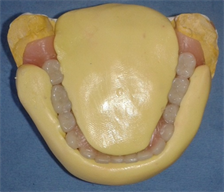
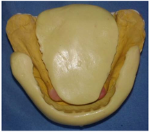
Diagnostic waxes were used to validate esthetics (Figure 2), labial support and occlusal vertical dimension, and to explain the prosthetic project to the patient. They were also used to make vestibular and lingual silicone matrix (Figure 3).
The purpose of these matrixes is to evaluate the available prosthetic space in the anteroposterior direction, taking as a reference the ideal profiles of the prosthetic extrados, and to study the possibility of incorporating the entire attachment system without interfering with the tongue and mandibular anterior teeth. Inter-arch and anteroposterior spaces were sufficient in this case.
![]()
Figure 1. Mounting on an articulator allowed the study of the available prosthetic space.
(a) (b)
Figure 3. (a): Silicone matrix was made using high viscosity elastomer; (b): Evaluation of the prosthetic space horizontally.
Following the clinical and radiological assessment, as well as the analysis of diagnostic wax models, the treatment decision was a conventional complete removable prosthesis in the maxilla and tooth-supported prosthesis with two Rhein 83 ball attachments on 33 and 43 in the mandible. The attachments comprise two elements: A male cast root cap with the retentive element and a corresponding female matrix incorporated into the fitting surface of the prostheses.
Both canines were scaled, and the patient was instructed in hygiene measures to ensure the optimal cleaning of the remaining teeth. Endodontic preparation was carried out followed by a tight sealing obturation of 33 and 43 (Figure 4).
The teeth were trimmed to a height of 1 to 2 mm above the gingival margin. The peripheral limit was in the form of a juxta-gingival peripheral chamfer. The occlusal opening was prepared in the form of an ovoid section funnel to contribute to stabilization (Figure 5).
The root preparation had a cylindrical-conical shape. Its length should occupy between 2/3 and 3/4 of the length of the root, and the width should not exceed one-third of the mesio-distal root width on the radiographic image to avoid weakening it. Apical sealing must be maintained by preserving a minimum of 3 to 5 millimeters of the apical filling.
The impression of the housing was made using the classic double-mix technique. A low viscosity silicone was injected with an appropriate syringe, then a plastic post was placed in the canal, and finally, the impression was taken with a tray loaded with high viscosity silicone (Figure 6). The two-step impression technique was not indicated in this case due to errors related to the repositioning of the impression made with a high-viscosity material on support surfaces.
![]()
Figure 4. Endodontic treatment of 33 and 43.
The primary impressions were made with plaster impression material. Root canal orifices should be protected with Vaseline-coated cotton balls (Figure 7). The impression was poured and custom trays were made using self-cure acrylic resin.
The copings with attachments were finished and polished after their manufacture in the laboratory and then tried in the patient’s mouth. The following were checked: Adaptation of the coping to the cervical margin, insertion, stability and primary retention.
Conventional techniques were employed to perform border molding using a thermoplastic impression material. The final impression was made using regular elastomeric impression material (Permlastic Regular), taking away the metal copings (Figure 8).
Once the final maxillary and mandibular impressions have been boxed and poured, occlusal models were made and the maxillo-mandibular relation was recorded. The choice of prosthetic teeth was made followed by teeth setting and try in (Figure 9).
After a satisfactory try-in, the final denture was cured using heat-cure acrylic resin. The prosthesis was tried with the attachments in place to check the spacing. Occlusal pressure must not cause any instability of the mandibular prosthesis.
Space and vent holes were created in front of abutment teeth (Figure 10). The copings with attachments were luted to the abutment teeth using glass ionomer cement (Figure 11). Occlusion was verified and then spacing was made with a small piece of glove (sheets of dental dam can also be used) (Figure 12). A self-curing resin was placed in the future location of the female part of the attachment, then the mandibular prosthesis was placed in the mouth. Upper and lower dentures were maintained in occlusion. Excess resin was eliminated and re-polishing was done. Then, an occlusion check and occlusal equilibration were performed (Figure 13).
The patient received guidance on the insertion and removal of the denture, along with instructions on eating and speaking. Maintenance of the results obtained on the day of placement is ensured by restoring good bucco-prosthetic hygiene and respecting periodic follow-up visits.
![]()
Figure 8. Mandibular secondary impression.
![]()
Figure 10. Space was created in front of abutment teeth.
![]()
Figure 11. Male parts were luted to the abutment teeth using glass ionomer cement.
![]()
Figure 12. Spacing was made with a small piece of glove.
![]()
Figure 13. The patient’s satisfaction smile after insertion of the prosthesis.
3. Discussion
Overdenture is a prosthesis that rests on both the entire bearing surface and the remaining roots to improve stabilization, support and retention [12] [13] [14] .
Overdentures are an interesting alternative to conventional complete removable prostheses, especially when additional retention systems are required. It allows to delayed progressive reduction of the residual ridge, improved prosthesis stability and makes mastication efficient [15] .
Abutment teeth play a crucial role in transmitting masticatory forces, thereby safeguarding the underlying mucosa and alveolar bone. Additionally, they contribute to maintaining proprioception, as the receptors of the periodontal ligament remain intact [7] .
Reducing the height of the abutment teeth to approximately 2 mm above the gingival margin reduces tooth mobility.
In the case of tooth loss, ODs can readily be converted into complete dentures [7] .
The indication for root-supported prosthetic treatment depends on a number of parameters. The distribution, number and type of teeth must be studied for their importance to prosthetic stability [1] . From a biomechanical point of view, the most favourable situation is to keep the teeth in symmetrical positions referred to the sagittal median axis. The most common situation is to retain either canines, or even both premolars [1] . Because of the length of their roots, their position in the arch, and their tendency to survive longest, canines are commonly selected as abutments for overdentures [15] . The number of residual roots must be less than or equal to 4 [1] .
The periodontal tissue of selected teeth should be free of inflammation, have a healthy, firm, pink, granulated appearance and present a sufficient band of attached gingiva measuring 3 - 4 mm. The depth of the vestibule should be free of deep undercut. Retained teeth must be free of periodontal pockets if they will be used for retention. A tooth with a maximum pocket of 4 mm will only be used for support [5] .
The bone tissue of residual roots must be evaluated. Indeed, reducing the height of the tooth helps to reduce the effects of mechanical stresses exerted by the prosthesis. The clinical root that will ensure a retention function must have sufficient amount of the bone surrounding the root that cannot be less than half of its morphological height. A minimum root length of 10 mm is desirable to indicate attachments. Canal preparation is done on two-thirds, keeping ideally 5 mm, and at least 3 mm of the apical filling [1] .
Diagnostic casts are used to validate the aesthetic and functional prosthetic project. Their analysis on an articulator enables us to detect the lack of space in the vertical direction, which can be a major contraindication to overdenture with attachment. Assessment of space in the vestibulo-lingual direction is also mandatory during the pre-prosthetic phase. A prosthetic space of 7 mm in height and 5 mm in width is sufficient for most attachment systems [1] . The presence of a pronounced undercut facing the abutment teeth may prevent the creation of a continuous prosthetic margin and therefore contraindicate the choice of overdenture as a treatment option [1] .
In this patient’s case, the pre-prosthetic study revealed the presence of sufficient prosthetic space for copings and axial attachments, and the absence of an anterior undercut.
In the case of overdentures with retention systems, prosthetic support must be provided entirely by the mandibular complete denture, without any involvement of the residual teeth, whose only function is to provide additional retention for the conventional complete denture. For this reason the mandibular complete prosthesis must be performed according to the classical requirements of the removable complete prosthesis [8] [16] .
Attachment systems must allow axial movement (vertical translation) and/or angular movement (distal rotation) to consider the difference in compressibility between desmodonts and fibromucosa. The attachment system must ensure retention through an articulated joint between the male and female components [1] .
Spaces allow for the development of a complete removable prosthesis that does not stress residual roots and their periodontal receptors in terms of support, thus eliminating any proprioception that could compromise the occlusal scheme of the complete prosthesis with mandibular displacements in para-centered or eccentric positions. These spacings are 0.2 to 0.3 mm for the gingival festoon and 0.5 mm for the copings and the top of the male parts [8] .
The periodontal health of retained roots can be maintained if the patient is motivated. Lack of motivation for oral-prosthetic hygiene is a contraindication to overdentures. Ball attachments are easy to brush. However, it is more difficult to maintain hygiene with bar attachment especially in elderly patients with reduced manual dexterity [1] .
Patients need to be instructed about the use of hygiene accessories, and how to properly clean their abutments and prostheses adequately at least twice a day. It is also crucial to educate them about the necessity of taking out their dentures when sleeping [15] .
It is essential to maintain and respect follow-up appointments, during which the clinician checks the occlusion and performs occlusal equilibration, checks the patient’s motivation for hygiene, checks the periodontal and fibromucosal condition of the bearing surfaces, and relines the prostheses.
Successful treatment depends on a number of factors, including the choice of abutment teeth, rigorous execution and mastery of techniques at every stage, and patient motivation.
4. Conclusion
Preserving dental roots is a viable option for improving the retention, stability and optimizing the prosthetic equilibrium of mandibular complete dentures. The use of various techniques to improve prosthetic stability and retention using attachments does not exempt the practitioner from rigorously carrying out the classic steps of complete denture. These must be performed with precision in order to maintain the retention systems and avoid possible therapeutic failure. Equally crucial is the patient’s awareness of the significance of follow-up visits, emphasizing the collaborative role between the practitioner and the patient in ensuring the long-term success of the prosthetic treatment.