Development of Preparation Method for Microencapsulating Uycalyptus Oil Containing Fine Aqueous Droplets by Use of Interfacial Condensation Reaction between Hydroxy Propyl Methyl Cellulose and Tannic Acid ()
1. Introduction
Many kinds of microcapsules have been developed and applied to the various fields such as cosmetics, paintings, catalyst, food industry, medicine, agriculture, and so on [1] -[3] .
The purposes of microencapsulation are to optionally release the core material, to protect the core materials from environment, to modify the surface of core material, to mask the taste of core material and so on. These functions of microcapsules are strongly dependent on the physical properties of shell and core materials, the structure, size and morphology of microcapsules. Accordingly, in order to prepare the microcapsules with the desired functions, it is necessary to develop the preparation method which is suitable to the physical properties of the core and shell materials used.
Recently, many studies about the microencapsulation of essential oils have been reported [4] -[10] . These microcapsules have been applied to the textiles, fragrance, aromatherapy, insect repelling, stress reducing, antibiosis and so on.
Uycalyptus oil is known to have many physiological activities such as anti-aging effect, anti-inflammation effect, sterilization effect, anti-uilus and so on. If the microcapsules containing the uycalyptus oil could be prepared and the oil could be optionally released, the uycalyptus oil will be utilized extremely easily in the more fields. Furthermore, it will be expected that the microcapsules containing the uycalyptus oil have to develop the new fields and the applicable fields of oil will be dramatically increased. Until now, many kinds of oil species have been microencapsulated with the chemical methods such as interfacial gelling reaction method [11] -[13] , the in-situ polymerization [14] [15] , the interfacial polycondensation reaction [16] [17] and the physicochemical methods such as the coacervation method [18] [19] , the spray-dried method [20] -[22] , and the melting dispersion-cooling method [23] . In these preparation methods, a few harmful chemical species have been used and the complicated processes have been applied. However, if we are going to apply the microcapsules to the cosmetics, the food and the drug, the microcapsules have to be prepared with the materials suitable to the living body and the edible materials. Accordingly, it is necessary to newly develop the preparation method by using the designnated materials with the simple process. The purposes of this paper are to develop the microencapsulation procedure with the interfacial condensation reaction between hydroxyl propyl methyl cellulose and tannic acid, to investigate whether the microcapsules of uycalyptus oil containing the fine water droplets can be prepared or not, to characterize the microcapsules and to discuss the microencapsulation mechanism on the basis of the results obtained.
2. Experimental
2.1. Materials
Materials used to develop the preparation method for microencapsulating the uycalyptus oil containing the fine water droplets are as follows:
The first core material was Uycalyptus oil (UO) (Wako Junyaku Co., Ltd.). Span 80 (Wako Junyaku Co., Ltd.) and Soybean Lecithin (Wako Junyaku Co., Ltd.) were used as the oil soluble surfactant. Hydroxy propyl methyl cellulose (HPMC) (50SH-50: Shinetsu Chemical Ind Co., Ltd.) and Tannic acid (TA) were used as the reactants to form the microcapsule shell. These chemical species were used as received.
2.2. Pre-Microencapsulation
In order to investigate whether the microcapsules can be prepared by the microencapsulation mechanism presented in this study or not, the pre-microencapsulation experiment was tried as follows.
Figure 1 shows the schematic diagram of pre-microencapsulation procedure. Namely, the HPMC aqueous solution and the UO were poured into the beaker as shown in Figure 1. The UO and the HPMC aqueous solution were separated to form the upper oil phase and the lower aqueous phase, respectively. Then, the TA aqueous solution was dropped into the oil phase through the nozzle by the syringe pump. If TA could transfer through the oil phase and react with HPMC on the interface between the oil phase and the HPMC aqueous solution, the gelated HPMC film should be formed on the interface. As a result, the TA aqueous droplet may be maintained on the formed film for a while.
According to this microcapsule shell formation mechanism, the pre-microencapsulation experiment was performed by changing the concentrations of HPMC and TA.

Figure 1. Pre-microencapsulation procedure.
2.3. Preparation of Microcapsules
Figure 2 shows the schematic diagram of experimental apparatus used to prepare the microcapsules. The reactor was the separable flask with the effective volume of 500 cm3. Four baffles made of aluminium plate were set on the wall of reactor. The six bladed disc turbine impeller with the diameter of 5 cm was used to stir the reaction mixture. The reactor was set in the thermos tatted water bath to keep temperature of reaction mixture constant. Figure 3 shows the flow chart for microencapsulating the UO containing the fine water droplets by using the interfacial condensation reaction between HPMC and TA. The aqueous solution dissolving TA as a gelation agent was dispersed into the UO dissolving Soybean Lecithin (SL) of an oil soluble surfactant to form the (W/O) emulsion.
Then, the (W/O) emulsion was dispersed into the continuous water phase dissolving HPMC to form the (W/O)/W emulsion. The operation stated above was performed at room temperature.
After formation of the (W/O)/W emulsion, temperature of the (W/O)/W emulsion was raised to 30˚C to perform the condensation reaction between TA and HPMC. When the reaction was continued for 1h, it was investigated whether the microcapsules of UO containing the fine water droplets could be prepared or not.
The formation of microcapsules was confirmed by observing the stability of the (W/O)/W emulsion after the microencapsulation process and by optical microscope. If the microcapsules were prepared well, the photographs of them were taken. Contrary to this, if the microcapsules were not prepared well, the emulsion should be broken rapidly.
In the fundamental experiment stated above, the concentrations of HPMC and TA were mainly changed. The experimental conditions in this experiment were shown in Table1
2.4. Characterization
2.4.1. Mean Diameter of Microcapsules
The microcapsules were observed by optical microscope and the photographs of them were taken. The diameter distributions and mean diameters of microcapsules were measured directly from these photographs.
2.4.2. Stability of Emulsion and Microcapsules
In order to investigate whether the microcapsules can be prepared or not, the (W/O)/W emulsion after microencapsulation process was set for 10 min. If the microcapsules could not be prepared well, the (W/O) droplets should be rapidly broken. The shape and inner structure of microcapsules were observed by taking the optical microscopic photographs.
2.4.3. Observation of Formation of Microcapsule Shell
The formation of microcapsule shell was observed to find the optimum concentrations of HPMC and TA.
Namely, the UO and the HPMC aqueous solution were poured into the beaker. The UO floated on the HPMC aqueous solution and the interface between the oil phase and the HPMC aqueous solution was formed as shown

Figure 2. Schematic diagram of experimental apparatus.

Figure 3. Flow chart for preparing microcapsules.

Table 1. Experimental conditions.
in Figure 1. The time elapsing from arrival of TA aqueous droplet at the interface to disappearance of droplet was measured. The concentrations of HPMC and TA required to form the gelated HPMC film could be estimated by observing the stability of a TA aqueous droplet on the interface.
3. Results and Discussion
3.1. Confirmation of Shell Formation and Microencapsulation Mechanism
Figure 4 shows the photographs of the TA aqueous droplet and the interface on which the gelated HPMC film was formed according to the concentrations of HPMC and TA. When the concentrations of HPMC and TA were low (CHPMC = 0.05 wt%, CTA = 0.01 mol), the TA aqueous droplet was kept on the interface as shown in Figure 4(a), but dissolved into the HPMC aqueous solution in a few min. The short life of TA aqueous droplet on the interface may be due to the formation of thinner film. On the other hand, when the concentrations of HPMC and TA were higher (CHPMC = 0.2 wt%, CTA = 0.06 mol), the TA aqueous droplet was kept on the interface for ca. 10 min as shown in Figure 4(b) due to the thicker film formation.
Figure 5 shows the measured results of the time elapsing from arrival of a TA aqueous droplet at the interface to disappearance. From these results, it is found that the film was not formed at CTA = 0 in spite of elapsing of ca. 30 s and the gelated film was formed above the concentrations of CHPMC = 0.05 wt% and CTA = 0.01 mol. Here, ca. 8 min at CHPMC = 0.05 wt%, and ca. 10 min at CHPMC > 0.1 wt% don’t mean that the TA aqueous droplet disappeared at these elapsing times, but mean that the measurement of elapsing time was finished at these times.
From these results, as the gelated HPMC film was found to be formed on the interface between the UO and the HPMC aqueous solution above CHPMC = 0.05 wt% and CTA = 0.01 mol. Taking these results into consideration, the formation mechanism of film may be stated as shown in figure 6. TA should transfer from the TA aqueous droplet to the interface through the UO and react with HPMC on the interface to form the gelated HPMC film. If the concentrations of HPMC and TA are lower, the thinner shell film should be formed. Contrary to this, if the concentrations of HPMC and TA are higher, the thicker and stronger shell film should be formed.
Furthermore, it was investigated whether the microcapsules could be prepared or not by the preparation method presented in this study. Namely, the stability of the (W/O)/W emulsion was observed with and without the microencapsulation process.
Figure 7 shows the photographs of the (W/O)/W emulsion just after the microencapsulation process (a) and after 10 min (b). The (W/O)/W emulsion without the microencapsulation process was separated rapidly into the oil phase and the water phase, but the observation result is not shown. Contrary to this, the (W/O)/W emulsion with the microencapsulation process, namely the (W/O) droplets microencapsulated floated on the water phase and was kept to stably disperse as shown in Figure 7(b). From these results, the UO droplets containing the fine water droplets were estimated to be microencapsulated well by the gelated HPMC shell.
3.2. Observation of Microcapsules
Figure 8 shows the optical microscopic photographs of microcapsules prepared by changing the concentration of HPMC at CTA = 0.06 mol. From these photographs, the following interesting results are obtained. The microcapsules could be prepared according to the formation mechanism presented in this study. The mean diameters of microcapsules changed from 780 µm to 1330 µm by changing the concentration of HPMC as shown in Figure 8. The fine water droplets were microencapsulated well, but become larger due to coalescence between the fine water droplets. In order to stably disperse the fine water droplets in the oil droplet and to utilize as the first core material, it must be necessary to select the optimum oil soluble surfactant species and the concentrations of them. The microcapsules become irregular with the concentration of HPMC. This result is considered to be due to the fact that the oil droplets containing the fine water droplets are hard to break by the stronger shell formed with the higher concentration and become irregular. These microcapsules could be stably dispersed in the continuous water phase under stirring of revolution speeds from 200 rpm to 500 rpm with the six bladed disc turbine impeller of 5 cm diameter.
3.3. Formation Mechanism of Microcapsules
From the results obtained above, the formation mechanism of microcapsules may be presented as shown in Figure 9. TA dissolved in the inner water droplets should transfer to the interface through the UO phase, react with HPMC on the interface and form the gelated HPMC film as the microcapsule shell.
In the case of the lower concentration of HPMC or TA, as the weak shell is formed, the (W/O) emulsion may be broken. As a result, the microcapsules cannot be prepared. Contrary to this, in the case of the higher concentration of HPMC and TA, as the stronger shell is formed, the microcapsules can be prepared well. The fine inner

Figure 4. Confirmation of microcapsule shell formation.

Figure 5. Effect of concentrations of HPMC and TA on film formations.
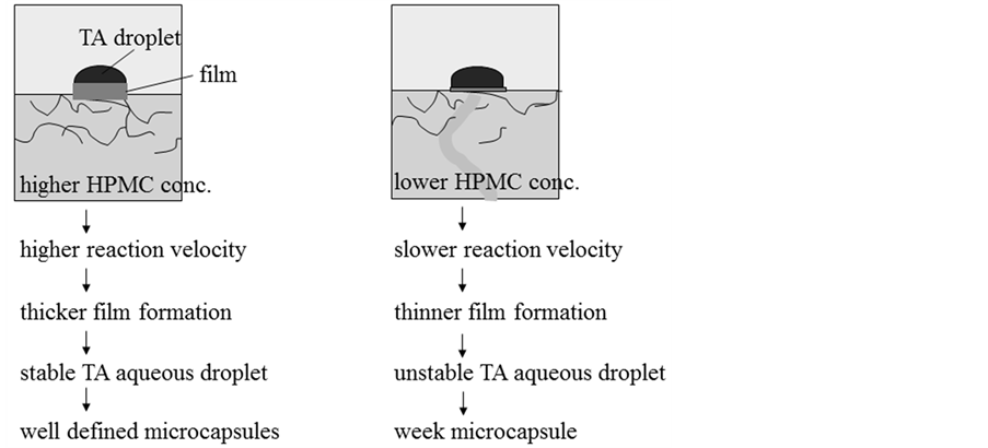
Figure 6. Formation mechanism of gelated HPMC film.
water droplets may coalesce each other to form the larger water droplets. If the fine water droplets could be dispersed stably, TA may transfer rapidly due to the larger interface area between the UO phase and the inner water phase. As a result, the shell may be formed rapidly and the microcapsules may be prepared more satisfactory. As many kinds of chemical species can be dissolved in the fine inner water droplets as the first core material and in the oil phase as the second core material at the same time, the microcapsules will be given the many kinds of functions. Namely, the microcapsules with the multiple functions can be prepared by using the microencapsulation method developed in this study.
However, it has to be investigated whether the microcapsules with the other oil species can be prepared by the microencapsulation mechanism presented here or not. Because, as it is necessary for TA to transfer through the
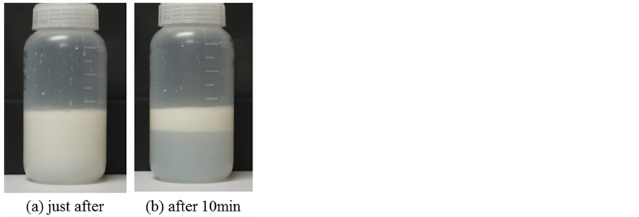
Figure 7. Photographs of (W/O)/W emulsion just after microencapsulation and after elepsing 10 min.

Figure 8. Photographs of microcapsules prepared by changing concentrations of HPMC.
oil phase, the fundamental experiments with regard to mass transfer of TA in the other oil species have to be performed.
4. Conclusions
It was tried to microencapsulate the uycalyptus oil droplets containing the fine water droplets according to the microencapsulation mechanism presented. The following results were obtained.
1) TA transferred from the water droplets to the interface between the uycalyptus oil and the HPMC aqueous solution through the uycalyptus oil and reacted with HPMC.
2) The preparation method for microencapsulating the uycalyptus oil containing the fine water droplets could be developed.
3) There were the critical concentrations (CHPMC = 0.05 wt%, CTA = 0.01 mol) for HPMC and TA required to prepare the sound microcapsules.
4) The microcapsules become irregular with the concentrations of HPMC.
5) The fine water droplets coalesced each other to form the larger water droplet.
6) As the microencapsulation method developed is the simple process with nontoxic materials, it may be expected that this method is going to be applied in many kinds of fields.
NOTES
*Corresponding author.