1. Introduction
This paper builds on an earlier model suggesting that quantum tunneling techniques which, in theory can be used successfully in cancer therapy [1]. This approach used the wave function of an electron to calculate the energy needed for an electron to “tunnel” through the cancerous membrane of the cell. It was suggested that this method could be more effective than conventional radiotherapy in treating cancer for the following reasons:
• In addition, the electron beam (which tunnels) can travel directly to the nuclei of the cancer cells.
• It is more direct, as instead of harming the surrounding cells it only targets the cancer cells through careful instrumentation and accuracy.
• According to the laws of momentum, there will be no impact from the electrons as they aim into the cells’ nuclei because if they tunnel, they must be travelling as a wave.
Nevertheless, [1] only suggests a hypothesis, however in this paper; we calculate the probability density of a single electron (in one dimension) tunneling into a cancer cell.
In this paper, we give accurately calculated figures of the probability density from the one-dimensional, time independent electron wave function, and we discuss the graph describing the electrons’ helical motion.
2. Quantum Wave Functions
Using the three parametric functions below, we combine them to create a helical graph which denotes the wave function, and hence the trail of an electron through space as a wave.
 (1)
(1)
 (2)
(2)
 (3)
(3)
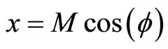
where x, y and z are the three Cartesian axes, M is the modulus of the helix, also equal to the amplitude of the helical wave. The parameter M is introduced to differentiate between wave functions of helixes of different amplitudes.  is the phase of the wave.
Given that:
 (4)
(4)
[2], we produce a graph of the parametric Equations (1)- (3).
In this case, the spiral will be no more than five times the diameter of the electron, therefore we can assume that . Using this figure, we can make calculations for the electron gun model [3].
3. Mathematical Modeling
3.1. Quantum Physical Properties of the Electron Wave
The process of mathematically modeling the electron wave function will find the realistic most efficient distance from the tumor by carrying out a triple integration of the wave function through a cancer cell (assuming that a cell is mostly H2O), to find a probability density of the electron tunneling into the cell [4].
We calculate the minimum and maximum energy required for the electron to be in waveform from Planck’s equations for the energy of a wave; E = hv [5], we calculate the minimum energy, which is determined by Equation (3) because of , the phase of the helix.
This equation leaves us with the simple relation between the frequency (f) and the phase ( ). As the coefficient of
  increases, the denominator of f divides by two.
 (5)
(5)
However, the frequency of the wave is a limit because although it will continue to approach zero, it will never reach, therefore, the definitions between particle and wave becomes fuzzy.
JB Hagen (2009) [6] suggested a different model for phase-angular frequency relation in electromagnetic waves; however, according to the laws of quantum mechanics, this is equivalent for electrons [7]. This model states that
 (6)
(6)
We now substitute into Planck’s equation for the energy of a wave, and we obtain the values:
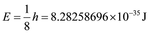 (7)
(7)
This is the minimum energy required for an electron to tunnel into the cancer cell. In addition, we can calculate the maximum energy that the electron can contain to be a wave.
As the electron wave-function verges towards the limit of a straight line rather than the helix graph, the behavior of the electron begins to be more particle-like, as it no longer can pass through “barriers”, and must give in to the laws of momentum. Einstein’s equation for the momentum of particles is the following: 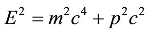 [8].
[8].
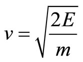
Therefore, we can calculate the electron’s momentum of impact on the cell wall. Given that the equation for kinetic energy is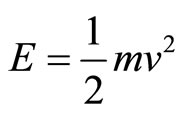 , we isolate v to then calcu-
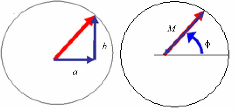
Figure 2. Cross-section view of the helix [2].
late Einstein’s equation for the momentum of a particle.
 (8)
(8)
 (9)
(9)
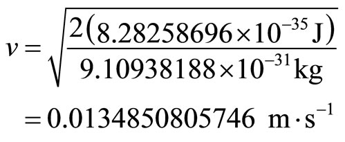
Therefore, by substitution, we calculate the velocity of the electron as 0.0134850805746 ms–1. Now we can calculate the momentum of the electron to be:

3.2. Probability Density
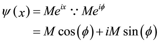
We calculate a one-dimensional probability density of an electron appearing “past the barrier” into the cancer cell. We obtain this figure from the one-dimensional wave function:  1 [2], which is an exponential function. We can therefore plot a 2D graph which represents our probability density function [9].
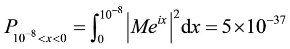
The integral which calculates the probability density function is calculated between two points, in this case, the diameter of a cancer cell [10].
 (10)
(10)
In this example, the integral would be:
 (11)
(11)
We calculate the integral from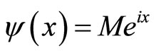 . M is the
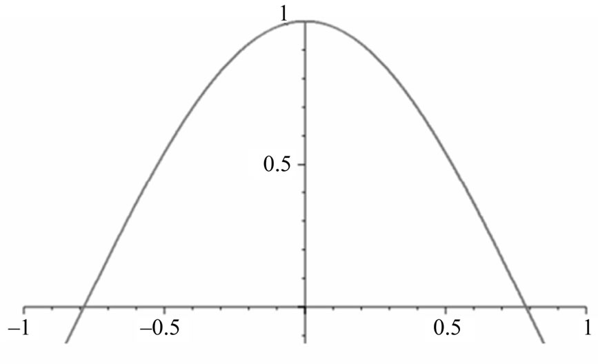
Figure 3. One-dimensional wave function graph for ψ(x).
modulus, the radius from the centre of the corkscrew to the limit .
.
We first normalize the equation into the form of a definite integral between ∞ and –∞.
 (12)
(12)
We now obtain a numerical value for the probability density distribution:
 (13)
(13)
4. Conclusion
We conclude this paper by providing numerical values for a probability density of a single electron tunneling into a cancer cell. In addition, we multiply this value by the number of electrons that we will shoot into a cancer cell, therefore increasing the electrons’ probabilities of tunneling. Given that in an electron beam with the width of 1 cm, there can be 5 × 10¹³ electrons, and our beam can be up to 10 cm long, we therefore calculate the volume of the beam (approximating a cubic prism) as being 10 cm3. In addition, we approximate that 1.25 × 1048 electrons can be within the space. According to the probability density which we have obtained, if we multiply our value of 5 × 10–37 by 1036 (the maximum number of electrons which we want to use) we obtain a probability of 0.5, which is a figure with a reasonable probability to allow electron wave therapy to commence.
5. Acknowledgements
We would like to thank Hannah Jones, Adam Biltcliffe and Professor Emanuele Giovannetti for constant support and fruitful discussions on the research.
NOTES