Open Journal of Obstetrics and Gynecology, 2011, 1, 197-201
doi:10.4236/ojog.2011.14038 Published Online December 2011 (http://www.SciRP.org/journal/ojog/ OJOG
).
Published Online December 2011 in SciRes. http://www.scirp.org/journal/OJOG
Conservative management of a recurrent puerperal uterine
inversion with bakri® balloon tamponade
Jesús Joaquín Hijona Elósegui1*, Francisco Javier Frutos Arenas1,2, Antonio Carballo García1,
Juan Antonio López López3, Gabriel Castilla Peinado4, Juan Manuel Torres Martí1
1Department of Obstetrics and Gynaecology, Complejo Hospitalario, Ciudad Real, Spain;
2Department of Radiology, Complejo Hospitalario, Ciudad Real, Spain;
3Department of Haematology, Complejo Hospitalario, Ciudad Real, Spain;
4Department of Anestesiology, Complejo Hospi talario, Ciudad Real , Sp ain.
Email: *jesushijona@gmail.com; jesushijona@gmail.com; javierfrutosarenas@yahoo.es; antoniogine@gmail.com;
juanantlop@yahoo.es; castillagabriel@hotmail.com; leonlennon@hotmail.com
Received 14 June 2011; revised 13 September 2011; accepted 29 September 2011.
ABSTRACT
Puerperal Uterine Inversion (PUI) is a rare but po-
tentially life-threatening delivery complicat ion in whi-
ch the uterine fundus collapses within the endome-
trial cavity. This “glove-finger” introflexion of uteri-
ne walls generally occurs as an immediate postpar-
tum complication and is responsible of different de-
grees of vaginal bleeding, shock and hypogastric pain
that can cause serious maternal complications, inclu-
ding death. There are few reports of recurrent post-
partum uterine inversion like the one we present here,
and its causes remain unclear. Early diagnosis of this
complication is crucial as it is the only one measure
that can allow a successful and conservative treat-
ment: an inverse relationship between the time that
uterus keeps inverted and the probability of reposi-
tioning has been firmly established. This case report
describes the exceptional and innovative use of the
SOS Bakri® balloon (Cook Medical Incorporated) in
the management of a recurrent puerperal uterine
inversion. To our knowledge it is one of the first re-
ports in the world of this procedure, perhaps the se-
cond one after Soleymani’s et al description; and the
first one in a third degree recurrent puerperal uteri-
ne inversion.
Keywords: Balloon Tamponade; Intrauterine; Manage-
ment; Postpartum Haemorrhage; Uterine Inversion; Re-
view; Treatm ent.
1. CASE REPORT
A 31-year-old healthy gravida 2, para 1 in established
labour presented at the delivery suite at 39 weeks’ + 5
weeks’ gestation. Her pregnancy had been uncomplica-
ted and all stages of labor progressed uneventful and
spontaneously, delivering a healthy 2400 gr male. Twen-
ty units of units of intravenous oxytocin were injected as
conventional early postpaturm haemorrhage prophylaxis.
One minute after delivery, the patient became restless.
She experienced severe pain and strong vaginal bleeding
started. She kept pushing and felt like bearing down a-
gain. Some second s later an elongu ed irregu lar and b loo-
dy mass protruded through the vulva and an unexpe-
cted finding of third degree puerperal uterine inversion
was diagnosed (Figure 1). Few seconds later the patient
was in hypovolaemic shock, pale and cold. At this mo-
ment her b lood pressur e was 85/50 mmHg and h eart rate
143 bpm .
Ressucitation maneuver were stablished inmediatly. A
facial mask oxygen was given (8 liters/minute) and other
18 G intravenous cannula was inserted. Intravenous col-
loid infusion commenced, a urinary catheter was inserted
and an attempt to replace the uterine inversion was made,
but this was not possible because of inadequate analgesia
and great cervical ring strength.
Figure 1. Acute third degree puerperal uterine
inversion. Note the elongued irregular and bl-
oody mass protruding through the vulva.
J. J. H. Elósegui et al. / Open Journal of Obstetrics and Gynecology 1 (2011) 197-201
198
The patient was transferred to theatre and an halo-
thane-based anaesthetic technique was performed. Com-
plete reduction was finally achieved by manual replace-
ment with Johnson maneuver (Figure 2) and the patient
was thoroughly examined to ensure that no other injuries
were sustained. At that point, the estimated blood loss
was about 1500 ml. After confirming successful repla-
cement of the uterus, which was now well contracted,
prophylactic intravenous antibiotics were administered
(1 gr ampiciline, 120 mg gentamicine and 500 mg me-
thronidazol), so as the administration of fourty units of
oxytocin and 0.250 mg of ergometrine maleate, in a 500
ml saline solution. Thirty minutes later, the patient beca-
me haemodynamically unstable again, with a blood pre-
ssure of 81/45 mmHg. At this time bleeding was poor
and vaginal examination confirmed that uterus had re-
inverted again showing fundus within vaginal cavity.
Once again the patien t was transferred to the treatre were
previously described procedures were repeated. At this
moment uterine fundus appeared atonic, and there was a
suspicion of great propensity to invert again (Figure 3).
As additional measure to prevent it a Surgical Obstetric
Silicone (SOS) Bakri® tamponade balloon catheter (Cook
Medical Incorporated) was inserted into the uterus with
Figure 2. Johnson manoeuvre: It consists of
pushing the inverted fundus through the cervi-
cal ring with pressure directed toward the um-
bilicus.
Figure 3. Repositioned uterus before Bakri®
balloon and vaginal packing insertion.
450 mL of normal saline solution to help preserving the
position of the fundus. A vaginal packing was also per-
formed. At that stage, the uterus was contracted and
there was no bleeding or abdominal pain. The patient
was transferred to a close puerperal observation area
from were she came back to the delivery suite 24 hours
later. Previously, the Bakri® balloon (Cook Medical In-
corporated) was deflated gradually and removed (oxyto-
cic and antibiotic infusion were kept during this proce-
dure). Recovery was unevetfull and patient was dischar-
ged home on day 3 with a short cou rse of or al antib iotics.
Any additional complication happened.
2. DISCUSSION
2.1. Definition
Puerperal Uterine Inversion (PUI) is a rare but poten-
tially life-threatening complication in which the uterine
fundus collapses within the endometrial cavity. This “glo-
ve-finger” introflexion of uterine walls [1] generally oc-
curs as an immediate postpartum complication and can
be either complete or partial.
Inversion of the uterus was more than likely first rec-
ognized by Hippocrates (460 a.C. - 370 a.C.) [2] but
other authors believe that Sorano (200 a.C.) was the first
one in describe this process and associating it with strong
traction on the umbilical cord [3].
2.2. Classification
Uterine inversion can be classified in some [2,4-6] de-
grees according to the intensity of introflexion. The most
extended classifications differences following types of
inversio n (Table 1):
There are few reports of recurrent PUI [7] like ours.
Causes of PUI remain unclear but there are two main
factors contributing: cervical dilation and smooth muscle
relaxation. That is why uterine inversion often happens
during the third stage of labour [8], particularly when a
strong cord traction is applied.
2.3. Incidence
Reported incidence of uterine inversion varies conside-
rably in literature from one in 8537 cases in Indian hos-
pitals to one in 27,902 ch ildbirths in British hospitals [9 ].
Van Vugt et al. reported 13 cases in 363,362 deliveries [4]
and Baskett reported puerperal inversion ranging from 1
in 1860 after caesarean section to 1 in 3737 vaginal de-
liveries [10].
2.4. Etiology
Even when the cause of uterine inversion remains un-
clear, several predisposing factors have been described
[11-12] (Table 2): Anyway, most cases are idiopathic
[13]. According to the previous, PUI has traditionally
C
opyright © 2011 SciRes. OJOG
J. J. H. Elósegui et al. / Open Journal of Obstetrics and Gynecology 1 (2011) 197-201 199
Table 1. Different classifications for uterine inversion.
According to the intensity of introflexion [2]
First-degree The inverted wall extends to (bu t not through) the cervix.
Second-degree The inverted wall protrudes through the cervix but remains within the vagina.
Third degree The inverted fundus extends outside t he vulva.
According to the existence or absence of tractions or other external str engths dur ing the third stage of labour[5]
Spontaneous
Secondary
According to the inverted portion [6]
Complete Inverted portion protrudes through the cervix.
Partial Inverted portion doesn’t protrude through the cervix.
According to the moment in which occurs considering delivery as a refere nce [4]
Acute Inversion occurs immediately after child birth.
Chronic Inversion occurs after a minimum of 30 days.
Subacute The inversion occurs between acute and chronic limits.
Table 2. Predisposing factors for uterine inversion.
DELIVERY RELATED FACTORS PATIENT RELATED FACTORS
Credé maneuver. Previous uterine inversion.
Low uterine tone. Uterine tumours and other anomalies.
Fundal insertion of placenta. Low parity.
Administration of oxytocin, particular ly when is admin is tered in bolus and
traction of the cord with the placenta, either partially or completely attached
to the uterus (adherent placenta). Young age.
Use of uterus-relaxing drugs. Inherent we akness of the uterine musculature and ligaments.
Cord shortness. Some authors12 suggest primiparity as a predisposing
factor, when it is associat e d to a fa st s ec on d st ag e
of labour, after a slow cer vi cal d ilat atio n.
Macrosomia.
High intraabdominal pressure.
been associated to “risky” interventions during the third
stage of labour. This belief is now being questioned due
to the poor incidenc e of this en tity, compared to the great
amount of deliveries in which cord traction and external
pressure over uterine fundus are applied [13-15].
Causes of few cases of recurrent inversion like the one
we present are even unclearer. It can be speculated that
some abnormalities of fundic myometrium may cause
ineffective retain [16] after uterine replacem ent.
2.5. Diagnosis
Is usually based on the presence of vaginal bleeding,
shock and hypogastric pain. Haemorrhage is the most
frequent symptom and shock seems to be secondary to
the blood loss and neurological response to the pelvic
ligamentarial traction [17]. Occasionally, when time per-
mits and equipment is urgently available, sonography
may help in diagnosis. Sonographic findings are striking
and can be easily understood if the pathologic process of
the uterine inversion is known. In the immediate post-
partum, the fluid-filled endometrial cavity must be easily
seen. Partial or complete inversion will result in poor
visualization of the fluid content, or in a Y-shaped con-
figuration caused by the invaginated fundus displacing
the two opposing uterine walls [18].
Early diagnosis of acute PUI allows a successful and
often conservative treatment, as there is an inverse rela-
tionship between the time uterus keeps inverted and the
probability of repositioning it as we previously signed
[19].
2.6. Tr eatment
Hypovolemia and shock must be immediate and appro-
piately corrected with crystalloids while aetiological treat-
ment should consist on manual manipulation of the ute-
rus to reposition it. Oxytocic agents must be avoided in
C
opyright © 2011 SciRes. OJOG
J. J. H. Elósegui et al. / Open Journal of Obstetrics and Gynecology 1 (2011) 197-201
200
this moment and sometimes other pharmacologic agents
can be employeed to assist uterine relaxation for achiev-
ing correction (tocolytic agents like terbutaline, magne-
sium sulphate and glyceryl trinitrate). If correction is not
obtained with tocolytic agents, general anaesthesia with
halothane may be induced to provide uterine relaxation.
This approach may be specially useful when the woman
is haemodynamically unstable, because halothane anae-
sthesia has fewer potential adverse effects on haemody-
namics [17,20]. About this concern, it is interesting to
note here that epidural analgesia does not help in uterine
reposition, as it does not affect uterine tone [21]. When
uterus is replaced, further agents are recommended to
achieve a powerful-enough uterine contraction able to
prevent uterine reinversion and decrease blood loss. If
these methods fail, surgical intervention might be nece-
ssary.
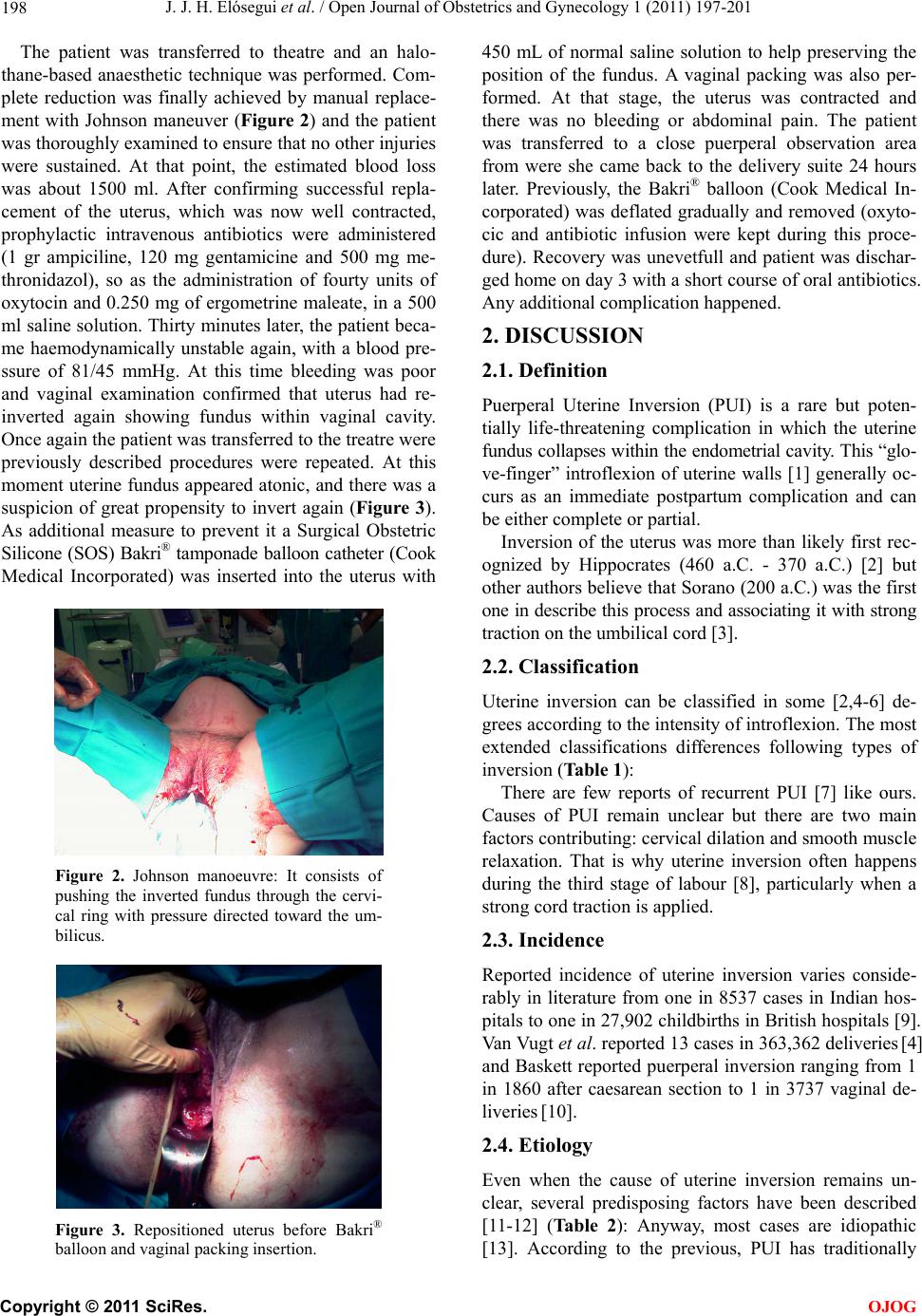
In our case, manual correction of uterine inversion was
made through the vagina by the Johnson maneuver, that
consists on pushing the inverted fundus through the cer-
vical ring with energic pressure directed toward the um-
bilicus [21,22]. It is generally suggested that removal of
the placenta before correction will result in increased
blood loss, so it is not recommended removing it, until
uterus has been replaced. Rates of immediate reduction
vary from 22% to 43% [17].
Apart from Johnson manoeuvre, some authors have
reported the use of hydrostatic pressure [23] caused by
warm water infused into the vagina to reduce uterine in-
version reduction.
When all attempts at manual reduction of the inver-
sion are unsuccessful, surgical correctionis often neces-
sary. The three most common procedures are the Hun-
tington, Haultain and Spinelli ones, but the first one is
considered the elegible. It consists on a laparotomy to
traction round ligaments and uterus to restore normal a-
natomy. Occasionally, as a life-saving measure, emer-
gency peripartum or obstetric hysterectomy is needed to
achieve control of haemorrhage [24].
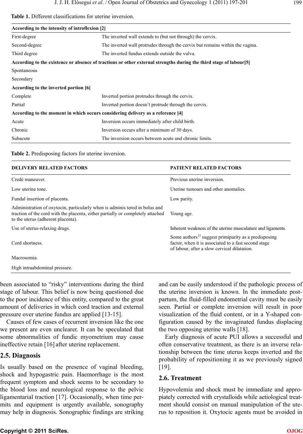
In our case surgical procedure was not necessary and
patient’s reproductive potential was kept, even being a re-
current case of uterine inversion. This conservative ma-
nagement of recurrent uterine inversion was based in
uterus reposition and insertion of a SOS Bakri® balloon
(Cook Medical Incorporated), which conformed to the
contours of the uterine cavity to prevent re-inversion of
the uterus.
The Bakri® tamponade balloon is 58 cm long silicone-
made inflatable balloon with a double lumen shaft, that
is easily removed transvaginally after deflation. Balloon
maximum capacity is 800 mL. and never should be in-
flated with less than 250 mL. However, the recommen-
ded use is up to 500 mL. The tip of the shaft has two
holes for drainage, so ongoing haemorrhage can be de-
tected after application of the balloon [25].
This case describes the exceptional and novedous use
of the SOS Bak ri® balloon (Cook Medical Incorporated)
in the management of recurrent puerperal uterine inver-
sion. To our knowledg e it is on e of the first reports in th e
world of this procedure, perhaps the second one after So-
leymani’s et al description [16] and the first one in a
third degree recurrent puerperal uterine inversion.
3. CONCLUSIONS
Insertion of intrauterine balloon tamponade may be an
effective measure to treat uterine inversion and prevent
its recurrence.
REFERENCES
[1] Morini, A., Angelini, R. and Giardini, G. (1994) Acute
puerperal uterine inversion: A report of 3 cases and an
analysis of 358 cases in the literature. Minerva Gine-
cologica, 46, 115-127.
[2] Siva, A., Zarina, M. and Mukudan, K. (2006) Puerperal
uterine inversion: A report of four cases. Journal of Ob-
stetrics and Gynaecology Research, 32, 341-345.
doi:10.1111/j.1447-0756.2006.00407.x
[3] Das, P. (1940) Inversion of the uterus. British Journal of
Obstetrics and Gynaecology, 47, 525-548.
doi:10.1111/j.1471-0528.1940.tb08843.x
[4] Van Vugt, P.J., Baudoin, P., Blom, V.M. and Van Deursen,
C.T. (1981) Inversio uteri puerperalis. Acta Obstetricia et
Gynecologica Scandinavica, 60, 353-362.
doi:10.3109/00016348109154123
[5] Milenkovic, M. and Khan, J. (2005) Inversion of the
uterus: A serious complication at childbirth. Acta Ob-
stetricia et Gynecologica Scandinavica, 84, 95-106.
doi:10.1111/j.0001-6349.2005.0254b.x
[6] Simó, M., Peñalva, G. and Domingo, X. (1992) Inversión
uterina obstétrica: Caso clínico y revisión del tema. Acta
Ginecológica, 49, 141-143.
[7] O’Connor, C.M. (1977) Recurrent postpartum uterine in-
version. British Journal of Obstetrics Gynaecology, 84,
789-790. doi:10.1111/j.1471-0528.1977.tb12494.x
[8] Bell, J.E. Jr., Wilson, G.F. and Wilson, L.A. (1953)
Puerperal inversion of the uterus. American Journal of
Obstettics Gynecology, 66, 767-780.
[9] Mehra, U. and Ostapowicz, F. (1976) Acute puerperal
inversion of the uterus in a primipara. Obstetrics and
Gynecology, 47, 30-32.
[10] Baskett, T.F. (2002) Acute uterine inversion. Review of
40 cases. Journal of Obstetrics and Gynaecology Canada,
24, 953-956.
[11] Vavilis, D., Tsolakidis, D., Goutzioulis, A.D. and Bontis,
J.N. (2008) Complete uterine inversion during caesarean.
Cases Journal, 1, 127. doi:10.1186/1757-1626-1-127
[12] Hostetler, D.R. and Bosworth, M.F. (2000) Uterine In-
version. A life-theatening Obstetric emergency. Journal
of the American Board of Family Practice, 3, 120-123.
[13] Parikshit, D.T. and Niranjan, M.M. (2004) Pregnancy
outcome after operative correction of puerperal uterine
inversion. Archives of Gynecology and Obstetrics, 69,
C
opyright © 2011 SciRes. OJOG
J. J. H. Elósegui et al. / Open Journal of Obstetrics and Gynecology 1 (2011) 197-201
Copyright © 2011 SciRes.
201
OJOG
214-216. doi:10.1007/s00404-002-0425-1
[14] Díaz, E., Paniagua, P., Segovia, O., Herrero, E., Díez
Gómez, E. and Guerra, J.M. (1994) Inversión uterina pu-
erperal: Caso clínico. Revisión de la literatura. Toko-Gin
Pract, 53, 313-316.
[15] Mohanty, A.K. and Trehan , A.K. (1998) Puerperal uterin e
inversion: Analysis of three cases managed by reposi-
tioning, and literature review. Journal of Obstetrics Gy-
naecology, 18, 353-354.
doi:10.1080/01443619867119
[16] Soleymani Majd, H., Pilsniak, A. and Reginald, P. (2009)
Recurrent uterine inversion: A novel treatment approach
using SOS Bakri balloon. British Journal of Obstetrics
and Gynaecology, 11 6, 999-1001.
doi:10.1111/j.1471-0528.2009.02165.x
[17] Beringer, R.M. and Patteril, M. (2004) Puerperal uterine
inversion and shock. British Journal of Anaesthesia, 92,
439-441. doi:10.1093/bja/aeh063
[18] Gross, R.C. and McGahan, J.I. (2009) Sonographic de-
tection of partial uterine inversion. American Journal of
Roentgenology, 144, 761.
[19] Ripley, D.L. (1999) Uterine emergencies. Atony, inver-
sion, and rupture. Obstetrics and Gynecology Clinics of
North America, 26, 419.
doi:10.1016/S0889-8545(05)70087-5
[20] Takeda, A., Manabe, S., Mitsui, T. and Nakamura, H.
(2006) Management of patients with ectopic pregnancy
with massive hemoperitoneum by laparoscopic surgery
with intraoperative autologous blood transfusion. Journal
of Minimally Invasive Gynecology, 13, 43-48.
doi:10.1016/j.jmig.2005.09.100
[21] Abouleish, E., Ali, V. , Joumaa, B., López, M. and Gupta,
D. (1995) Anaesthetic management of acute puerperal
uterine inversion. British Journal of Anaesthesia, 75,
486-487.
[22] Calder, A.A. (2000) Emergencies in operative obstetrics.
Best Practice and Research Clinical Obstetrics and Gy-
naecology, 14, 43-55. doi:10.1053/beog.1999.0062
[23] Vijayaraghavan, R. and Sujatha, Y. (2006) Acute post-
partum uterine inversion with haemorrhagic shock:
Laparoscopic reduction: A new method of management?
British Journal of Obstetrics and Gynaecology, 11 3,
1100-1102. doi:10.1111/j.1471-0528.2006.01052.x
[24] Sumera, T., Mahmood, A. and Samina, A. (2003) Indica-
tion and maternal outcome of emergency peripartum
hysterectomy. Pakistan Journal of Medical Sciences, 19,
182-186.
[25] Vitthala, S., Tsoumpou, I., Anjum, Z.K. and Aziz, N.A.
(2009) Use of bakri balloon in post-partum haemorrhage:
A series of 15 cases. The Australian and New Zealand
Journal of Obstetrics and Gynaecology, 49, 91-94.
doi:10.1111/j.1479-828X.2009.00968.x