Surgical Science, 2011, 2, 262-268
doi:10.4236/ss.2011.25058 Published Online July 2011 (http://www.SciRP.org/journal/ss)
Copyright © 2011 SciRes. SS
The Q-Switched Nd: YAG Laser in Tattoo Removal and the
Effect of Lymphatic Elimination: An Experimental
Study in Rabbits
S. Irkoren, C. Demirdover, B. Z. Akad, M. Gorgu
Adnan Menderes Universitesi, Tıp Fakültesi Plastik, ve Rekonstrüktif Cerrahi Anabilim Dalı
E-mail: saimeirkoren@hotmail.com
Received March 28, 20 1 1; revised June 20, 2011; accepted June 30, 2011
Abstract
Background: Previously described methods for removal of tattoos are chemical, mechanical, surgical, termal
and laser assited methods. Invention of the Q-switch mode, advanced the laser method, to be one of the most
effective methods of tattoo removal. Objective: Comparing the tattoo removal and rate of lymphatic elimina-
tion of 3 different wavelengths of Q-switched Nd-YAG laser (532nm, 1064nm, combination 532 + 1064).
Methods: In this study we examined lymphatic elimination and the effect of 532 nm, 1064 nm, and the com-
bination (532 + 1064 nm) wavelength of laser pulse for the possible lymphatic cleansing mechanism of black
pigmented tattoos. This study was performed on 18 New-Zealand rabbits, black pigmented tattoos were en-
graved on the back and the four extremities of the animals. 532 nm wavelength of Q switched Nd: YAG la-
ser beam was applied on the left upper and bilateral lower extremities of the rabbits. During this period, ex-
cisional skin biopsies and lymph node biopsies were performed on days 7, 14 and 21. Results: Day 21 lymph
node biopsies revealed mixed type of reactive hyperplasia and intracellular pigments were markedly seen in
the laser treatment group and no intracellular tattoo pigment was seen in the control group. Conclusion: The
findingd of this study indicate that lymphatic elimination may be one of the significant mechanisms of tattoo
removal and the application of different wavelengths of Q-switched Nd-YAG laser do not show statistically
significant differences in tattoo removal.
Keywords: Q-Switched Nd-YAG Laser, Tattoo Removal, Rabbits
1. Introduction
Traumatic and elective tattoo applications are wide-
spread in the society. People, either due to psychosocial
reasons or due to the constraint of society on them, want
their tattoos removed for many reasons. Studies reveal
that 42% of people apply to medical centers for the re-
moval of their tattoos sometime during their lives [1].
Removing tattoos has gained wide practice among
plastic surgeons. It is well known that pigment of tattoo
can migrate to the regional lymph nodes [1,2]. The car-
bon particles mobilize and migrate through the lymphat-
ics similar to melanoma cells and can be seen within the
histiocytes or extracellularly. However, in this study we
tried to show lymphatic eliminatio n of the tattoo pig ment
occuring during late post-interventional periods when
Q-switched Nd: YAG laser was utilized for treatment.
The aim of this study was to determine the timing of
lymphatic elimination of tattoo pigment after laser treat-
ment.
A variety of methods are used for removing tattoos
such as chemical, mechanical, surgical, thermal and laser.
However each method presents with its own advantages
and disadvantages. “Selective photothermolysis” theory
mentioned by Anderson and Parrish summarize that la-
sers only effect the pigment that are charged with wave-
length specifi c ch r omophores [ 3 -5] .
Therefore, the dermis and dermal appendages, whereby
the tattoo pigment lies, is protected so that the scar tissue
does not form with this method. Several types of lasers
such as Q- switched ruby laser, alexandrite laser and Nd:
YAG lasers which can transfer large amounts of energy
to the tissue in a matter of n anoseco nd s, are used in ord er
to remove tattoos, [6,7]. Th e effect of these laser types is
discussed in many studies bu t the “perfect tattoo laser” is
not invented to date [8-10]. As black is the most com-
S. IRKOREN ET AL.263
monly used tattoo pigment, we used Q-switched Nd:
YAG laser in different wavelengths including 532 nm,
1064 nm, and their combination (532 + 1064 nm) in our
study. Many studies in the literature have shown the ef-
fectivity of lasers in tattoo removal. However, the me-
chanism of action remains an area that is not agreed.
[11-13]. Studies have shown that lasers increase transe-
pidermal migration by fragmenting the pigments with
their photoacustic effects [14]. Also with their photother-
mal effects pigment are burned eventually causing fibro-
sis. [15]. In this study, the lymphatic elimination of tat-
too pigment after the application of 532 nm, 1064 nm
and 532 + 1064 nm wav elength of Q-switch ed Nd: YAG
laser was investigated on black pigmented tattoos applied
to 18 New-Zealand rabbits.
2. Materials and Method
Albino, male 18 New Zealand rabbits weighing between
1500 - 2000 grams are utilized for this study. Animals
were sedated with 1mg/kg midazolam (Dormicum®).
The extremities were shaved and EMLA cream was ap-
plied for 1 hour before the tattooing process was initiated
The tattoo gun, was a custom made 20 turns/second with
3 needles of 0.5 cm tips that has the ability to inject the
pigment 1 mm deep into the dermis. The tattoo pigment
was carbon based “Diamond Black” (CI 1253) and in-
dian ink as a mixture. A total of 48 tattoos were applied
on the extremities of the rabbits in a single session.
At the end of three weeks, laser treatment of different
wavelengths (532 nm, 1064 nm, and 532 + 1064 nm)
was applied to each animal and afterwards macroscopic
and histopathological observations were made as designed.
Animal model: Tattoos were drawn on the four ex-
tremities of each rabbit. Right upper extremity of the
animals were regarded as the control group. The left up-
per extremity and the bilateral lower extremities were
selected as the experimental group. There were three
experimental groups each including six rabbits (Table 1 ).
Groups were identified as Group 532, Group 1064 and
Group Combination (Scheme 1). Two passes of 532 nm,
1064 nm, and (532 + 1064) nm Q switched Nd: YAG
laser was applied on the extremities of the animals with
450 mJ energy, 3mm spot diameter, 2 Hz frequency.
Laser was applied to the extremities (Lightage, Q-Clear
Table 1. Design of experimental groups.
Gr-532 Gr-1064 Gr-532 + 1064 Gr-Control
Day 7 6 6 6 6
Day 14 6 6 6 6
Day 21 6 6 6 6
Scheme 1. Scheme representing the localization of lymph
node biopsies taken from group 532, group 1064, and group
combined (532 + 1064 nm).
®) in a single session. Skin and lymph node biopsies
were dissected on days 7, 14 and 21 (Figures 3-5). Axil-
lary and inguinal node dissection with skin islands was
performed as excisional biopsies. And Photographs were
taken for macroscopic assessment (Figures 6, 7). Mac-
roscopic assessment w as made with Nikon Coolpix 5000
5.0 megapixel camera digitally in order to subjectively
identify the amount of tattoo removal achieved with or
without laser therapy. Photographs taken of the tattoos
prior to and 6 weeks after treatment and were scored by
three independent blind observers. Each observer was
unaware of the study protocol and evaluated the tattoos
on a clinical scale of 0 - 4 (0 = “no change”, 1= “1% -
25% lightening, 3 = ”51% - 75% lightening and 4= “76%
- 99% lightening”) .
The animals were sacrified on the 21st day.
For histopathological assessment, microscopic slides
were dyed with hematoxylene eosine and gomori
trichrome dye and were examined under 20×, 40×, and
100× light microscopic magnification.
3. Results
Control group, showed signs of fading over time, how-
ever no lymphatic cleansing of the tattoo pigment was
noticed. This group was graded as “0% - 25% - poor
removal of tattoos”. All of the therapy groups showed
cleansing of the tattoo pigments to various extent (Fig-
ures 1, 2). Clinical scale of all treatment groups (scale 3)
were significantly better than the co ntrol group (scale 1).
Histopathological examination revealed no significant
difference between treatment groups (p > 0.05).
Copyright © 2011 SciRes. SS
S. IRKOREN ET AL.
264
The results of microscopic examination for each group
in various days is presented in Table 2.
Lymphoid hyperplasia in dissected nodes started to be
detected on 14th d ay. On 14th day, lymphoid clearing was
started in 4 animals in group 532-1064 and in 5 animals
in group 532 + 1064. On the 21st day, lypmphoid clear-
ance was detected in all animals in the treatment g roups.
In no lymphatic clearence was detected in the control
groups (Table 3).
4. Discussion
Laser removal of tattoos is based on the principle of se-
lective absorption. The tattoo pigment, which is the.
Figures 1. Macroscopic view of the tattoo on the extremity.
Figures 2. Macroscopic view of the cleansing after 532 nm
wavelength Q Switched Nd:YAG laser therapy.
Figures 3. Post-laser seventh day skin biopsies stained in
Haematoxylene-Eosine showing thermal necrosis and in-
flammatory cell reaction.
Figures 4. Post-laser seventh day skin biopsies stained in
Gomori-Trichrome showing thermal necrosis and inflam-
matory cell reac ti on.
Figures 5. Post-laser 21st day biopsies stained with Haema-
toxylene-Eosine (×20) showingmixed type lymphoid reaction.
Copyright © 2011 SciRes. SS
S. IRKOREN ET AL.
Copyright © 2011 SciRes. SS
265
Figures 7. Intralymphatic view of tattoo pigments in Go-
mori Trichrome (×100).
chromophore, absorbs the electromagnetic energy, the
resultant thermaleffect being confined to the pigment
The duration of exposure determines the extent of the
local heating effect. The thermal relaxation time is the
t1/2 time for tissue cooling and limiting exposure to less
than this minimises the thermal effects on the surround-
ing tissues. This concept is one of the rationales for
Qswitching a laser as it enables a high energy nanosec-
ond duration pulse to be delivered. The bulk of tattoo
Figures 6. Intralymphatic view of tattoo pigments in
Haematoxylene-Eosine (×100).
Table 2. Histopathological results comparing group-532, group-1064, group-532 + 1064, and group-control.
Gr-532 Gr-1064 Gr-532 + 1064 Gr-Control
Day 7
crusts of serous leakage
dermoepidermal junction is
intact
Moderate inflammatory
reaction of macrophages
and leucocytes each
containing tattoo pigments
crusts of serous leakage
dermoepidermal junction
is intact
Moderate inflammatory
reaction of macrophages
and leucocytes each
containing tattoo pigments
crusts of serous leakage
dermoepidermal junction
is intact
Wide-spread inflammatory
reaction of macrophages
and leucocytes each
containing tattoo pigments
extracellular pigments
localized in the
superficial dermis
Day 14
moderate inflammatory
reaction in the dermis
intracellular pigment
localization (phagocytosed
by the macrophages.
severe inflamm atory
reaction in the dermis
intracellular pigment
localization (phagocytosed
by the macrophages.
severe inflamm atory
reaction in the dermis
intracellular pigment
localization (phagocytosed
by the macrophages.
extracellular pigments
localized in the
superficial dermis
eosinophilically rich
dermis and mild
inflammatory reaction.
Day 21
Gomori Trichrome dye
revealed no signs of
fibroblast increase nor
fibrosis formation
In the histopathologica l
lymph node examination,
mixed type of reactive
hyperplasia and intracellular
pigments were seen
Gomori Trichrome dye
revealed no signs of
fibroblast increase nor
fibrosis formation
In the histopathologica l
lymph node examination,
mixed type of reactive
hyperplasia and
intracellular pigments were
seen
Gomori Trichrome dye
revealed no signs of
fibroblast increase nor
fibrosis formation
In the histopathologica l
lymph node examination,
mixed type of reactive
hyperplasia and
intracellular pigments
were seen
Pigments are
extracellularly localized
at the superficial dermis
Gomori Trichrome dye
revealed no signs of
fibroblast increase nor
fibrosis formation
In the histopathologica l
lymph node
examination,
intracellular pigments
were not seen
S. IRKOREN ET AL.
Copyright © 2011 SciRes. SS
266
Table 3. Isolation of the tattoo pigments on lymph node
biopsies.
Gr-532 Gr-1064 Gr-532 + 1064 Gr-Control
Day 7 0 0 0 0
Day 14 4 4 5 0
Day 21 6 6 6 0
pigment is found clumped within the dermis contained
within fibroblasts and macrophages [3,25]. Following
exposure, the production of CO 2 and steam in the dermis
forms vacuoles and causes the skin to whiten. This partly
accounts for the disappearance of the pigment but also
the pigment is disrupted into smaller particles allowing
phagocytosis over the ensuing weeks. The frequency
doubled Q-switched Nd: YAG laser (532 nm/1064 nm)
is the most efficient laser for clearing red, brown, orange
and black pigments [3,8]. As selective absorption by
pigment is partly wavelength dependent [3,24]. Different
kinds of ablative and nonablative lasers have been used
for tattoo removal. After the innovation of Q-switched
(QS) mode lasers, QS Nd: YAG, QS- Ruby, QS- Alex-
andrite lasers have been popularized in clinical use.
Apart from the Q-switched mode Nd: YAG lasers, CO2
lasers and ablative lasers are also used for “difficult to
remove” tattoos and tattoos composed of allergenic pig-
ments. Q-switched mode Nd: YAG laser, transfer large
amounts of energy to the epidermis in a matter of nano-
seconds [3,16]. Tattoo clearing may occur via particle
fragmantation, phagocytosis of cell death and subsequent
elimination via lymphatics [17,18]. Selective photothe-
molysis theory popularized by Anderson and Parrish
showed that Q-switch mode lasers show selective effects
for pigment retaining cells. With this method, injury to
the surrounding tissues is avoided by targeting only pig-
ment including cells [19-21]. In this study; we evaluated
lymphatic elimination with application of 532 nm, 1064
nm, and (532 + 1064) nm wavelengths of laser Also the
timing and initiation of lymphatic elimination was tried
to be established through serial biop sies.
Microscopically, laser therapy groups showed ondula-
tion in dermoepidermal junction and epidermal pigment.
containing keratinocytes. Studies in literature show that
during the application of the tattoo pigment, trauma
caused by the movements of the needle produce der-
moepidermal lacerations leading to pigment leakage
from this region which are then phagocytosed by kerati-
nocytes and removed from the dermis [3,22]. This period
lasts 7 - 10 days and ends when dermoepidermal unity is
preserved again. This mechanism is named “transdermal
migration” and is thought to be responsible for 30%
cleansing of the tattoo pigment. In our study this is the
mechanism of tattoo fainting in the control group after 1
month. However in laser therapy groups, pigment in
epidermal keratinocytes, increase laser’s effect on trans-
dermal migration. This may be due to basal membrane
injury by direct effect, leading to disintegration of basal
membrane cells by photoacustic effect and forming der-
moepidermal tunnels by photothermal effect. However,
electron microscopic studies are needed for further in-
vestigation.
Other dermal changes included thermal necrosis re-
gions superficially and empty vacuoles around these ne-
crotic regions. Pigments in laser therapy groups are
found to be placed as small fragments around the vacu-
oles, partially phagocytosed by the macrophages. Litera-
ture review reveals that these vacuoles are formed due to
the gas released after the thermal effect leading to in-
flammation reaction due to the photothermal affect [4].
These vacuoles are also hold responsible for the sudden
paleness of the tissues right after the administration of
the tattoo pigment. CO2 gas formed during the healing
procedure of carbon based dye [5,13]. Small scattered
fragment of pigments around the vacuoles are the results
of high amounts of energy exploding due to the photo-
acustic effect of lasers according to our study. Vacuoles
are found to disappear around day 7 and we believe that
the mechanism for this is due to gas filled cavities are
replaced by intertitial fluids. When comparing the con-
trol group with the laser therapy groups, the distribution
of pigment in the dermis is one of the most striking dif-
ferences. Previous studies revealed that photoacustic
effect of laser cause the larger fragments to disintegrate
and be easily phagocytosed by macrophagesThis may
explain the faster removal of dermis in treatment group.
[12]. Macrophages and polymorphonuclear leucocytes
are much greater in number in laser therapy groups due
to their role in effective phagocyto sis and removal of the
tattoo pigment. Some studies revealed that the tattoo
pigment remain in the fibroblast in long-term periods
[21]. In our study, none of the fibroblasts contained pig-
ment in any of the biopsies however this may be inter-
preted as more time (more than 6 weeks) may be needed
for that observation. Microscopic slides with Gomori
Trichrome, revealed less fibrosis in the laser therapy
groups, and this may be explained by selective injury of
lasers to the pigment containing cells.
The effect of lymphatic system in tattoo removal is a
subject discarded in many reports.
Some studies suggest a possible mechanism however,
no experimental studies have been performed on this
mechanism [7,10]. Tattoo is known to cause lymphade-
nopathy in the acute phase [2, 22]. This is thought to be
due to local inflammation from the initial insult. How-
ever tattoo pigment in the lymph nodes after 3 months
S. IRKOREN ET AL.267
can not be described as acute inflammation. In our study
we waited for 3 weeks for the acute inflammatory reac-
tion to setle and then we applied the laser beam.
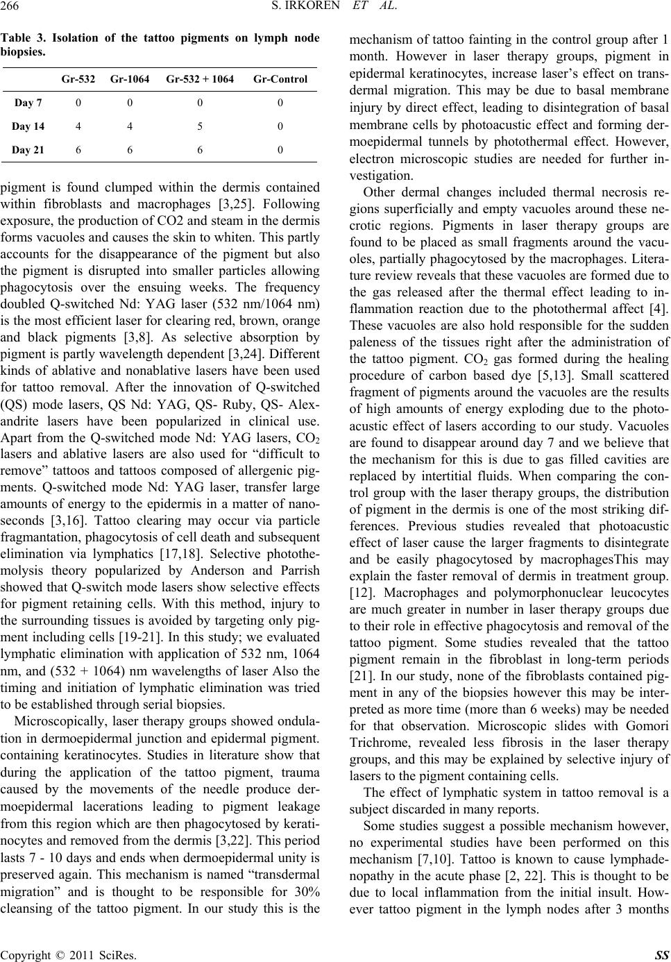
Our axillary and inguinal dissection results reveal a
possible elimination via the lymphatic system starting
from the 14 day post-laser treatment and makes a peak
around 21 days post-laser treatment lymph node biopsies.
Pigment that is fragmanted into small particles by laser
therapy migrates to the dermis, where they are carried to
the lymph nodes with protein enriched extracellular flu-
ids. Pigment in the lymphoid sinuses are phagocytosed
by macrophages and B,T cell lymphatics. This may initi-
ate the allergic reaction to some types of tattoos by po-
tentiating cellular and humoral immune responses [11].
The limitations of this study are several. First of all,
although we are unable to control the application and
type of pigment used in clinical practice. However, this
study is limited to black carbon pigments. The lymphatic
elimination may be further analyzed with other tattoo
pigments than the carbon molecules in future studies.
Secondly, in clinical practice we treat each tattoo 4 - 12
times for maximum lightening and elimination. In this
study, we evaluated the response after only a single dose
of 532 nm, 1064 nm and combination 532 + 1064 nm
wavelength Q-switched Nd: YAG laser treatment. We
also noticed no statistically significant difference be-
tween the different wavelengths of lasers in changing the
rate of lymphatic elimination.
5. Conclusions
As a result of this study we want to specifically empha-
size that lymphatic elimination starts to guide laser tattoo
treatment 14 days after a single application of 532 nm,
1064 nm and combination 532 and 1064 nm wavelength
Q-switched Nd: YAG laser treatment, with statistically
significant difference on cleansing rate of the tattoo pig-
ments, however further electron microsopic examination
regarding the role of lymphatic elimination in long term
results of laser treatment may be the inspiration for fu-
ture experimental studies.
6. Acknowledgements
We would like to thank Laserium corporation for pro-
viding us the Lightage Q-Clear Laser. Th eir contribution
is greatly valued.
7. References
[1] C. Jack, A. Adwani and H. Krishnan, “Tattoo Pigment in
an Axillary Lymph Node Simulating Metastatic Malig-
nant Melanoma,” International Seminars In Surgical
Oncology, Vol. 1, No. 2, 2005, p. 28.
doi:10.1186/1477-7800-2-28
[2] T. Friedman, M. Westreich, S. N. Mozes, A. Dorenbaum
and O. Herman, “Tattoo Pigment in Lymph Nodes Mim-
icking Metastatic Malignant Melanoma,” Plastic and
Reconstructive Surgery, Vol. 111, No. 6, 2003, pp. 2120-
2122. doi:10.1097/01.PRS.0000057101.95872.A1
[3] B. D. Zelickson, D. A. Mehregan, A. A. Zarrin, C. Coles,
P. Hartwig, S. Olson and J. Leaf-Davis, “Clinical, His-
tologic and Ultrastructural Evaluation of Tattoos Treated
with Three Laser Systems,” Lasers in Surgery and Medi-
cine, Vol. 15, No. 4, 1994, pp. 364-372.
doi:10.1002/lsm.1900150406
[4] D. M. Ho, R. London, G. B. Zimmerman and D. A.
Young, “Laser-Tattoo Removal—A Study of the Mecha-
nism and the Optimal Treatment Strategy via Computer
Simulations,” Lasers in Surgery and Medicine, Vol. 30,
No. 5, 2002, pp. 389-397. doi:10.1002/lsm.10065
[5] J. E. Ferguson, S. M. Andrew, C. J. P. Jones and P. J.
August, “The Q Switched Neodymium: YAG Laser and
Tattoos: A Microscopic Analysis of Laser-Tattoo Interac-
tions,” British Journal of Dermatology, Vol. 137, No. 3,
1997, pp. 405-410.
doi:10.1111/j.1365-2133.1997.tb03747.x
[6] M. L. Leuenberger, M. W. Mulas, T. R. Hata, M. P.
Goldman, R. E. Fitzpatrick and J. M. Grevelink, “Com-
parison of the Q-Switched Alexandrite, Nd: YAG, and
Ruby Lasers in Treating Blue Black Tattoos,” Derma-
tologic Surgery, Vol. 25, No. 1, 1999, pp. 10-14.
doi:10.1046/j.1524-4725.1999.08122.x
[7] J. E. Ferguson and P. J. August, “Evaluation of the Nd:
YAG Laser for Treatment of Amateur and Professional
Tattoos,” British journal of dermatology, Vol. 135, No. 4,
1996, pp. 586-591.
doi:10.1111/j.1365-2133.1996.tb03836.x
[8] S. L. Kilmer and R. R. Anderson, “Clinical use of the
Q-Switched Ruby and the Q-Switched Nd: YAG( 1064
nm and 532 nm) Lasers for Treatment of Tattoos,” Jour-
nal of Dermatologic Surgery & Oncology, Vol. 19, No. 4,
1993, pp. 330-338.
[9] S. L. Kilmer, M. S. Lee, J. M. Grevelink, T. J. Flotte and
R. R. Anderson, “The Q-Switched Nd: YAG Laser Effec-
tively Treats Tattoos. A Controlled, Dose Response Stu-
dy,” Archives of Dermatology, Vol. 129, No. 8, 1993, pp.
971-978. doi:10.1001/archderm.129.8.971
[10] S. E. Dozier, D. G. Diven, D. Jones, et al., “The Q-Swi-
tched Alexandrite Laser’s Effects on Tattoos in Guinea
Pigs and Harvested Human Skin,” Dermatologic Surgery,
Vol. 21, No. 3, 1995, pp. 237-240.
[11] R. O. Gregory, “Overview of Laser in Plastic Surgery,”
Clinics in Plastic Surgery, Vol. 25, No. 1, 1998, pp. 6-10.
[12] R. O. Gregory, “Laser Physics and Physiology,” Clinics
in Plastic Surgery, Vol. 25, No. 1, 1998, pp. 89-93.
[13] E. V. Ross, S. Yashar, N. Michaud, R. Fitzpatrick, R.
Geronemus, W. D. Tope and R. R. Anderson, “Tattoo
Darkening and Nonresponse after Laser Treatment. A
Possible Role for Titanium Dioxide,” Archives of Der-
matology, Vol. 137, 2001, pp. 33-37.
Copyright © 2011 SciRes. SS
S. IRKOREN ET AL.
Copyright © 2011 SciRes. SS
268
[14] B. E. DiBernardo and A. Cacciarelli, “Cutaneous Lasers,”
Clinics in Plastic Surgery, Vol. 32, No. 2, 2005, pp. 141-
150. doi:10.1016/j.cps.2004.11.008
[15] K. R. Suchin and S. S. Greenbaum, “Successful Treat-
ment of a Cosmetic Tattoo Using a Combination of La-
sers,” Dermatologic Surgery, Vol. 30, No. 1, 2004, pp.
105-107. doi:10.1111/j.1524-4725.2004.30024.x
[16] G. Jimenez, E. Weiss and J. M. Spence r, “Multiple Color
Changes Following Laser Therapy of Cosmetic Tattoos,”
Dermatologic Surgery, Vol. 28, No. 2, 2002, pp. 177-179.
doi:10.1046/j.1524-4725.2002.00161.x
[17] A. Jones, P. Roddey, I. Orengo and T. Rosen, “The
Q-Switched ND: YAG Laser Effectively Treats Tattoos
in Darkly Pigmented Skin,” Dermatologic Surgery, Vol.
22, 1996, pp. 990-1001.
[18] S. L. Kilmer, “Laser Treatment of Tattoos,” Lasers in
Dermatology, Vol. 15, No. 3, 1997, pp. 409-417.
[19] R. R. Anderson, “Regarding Tattoos,” Archives of Der-
matology, Vol. 137, 2001, pp. 210-212.
[20] C. Gundogan, B. Greve, I. Hausser and C. Raulin,
“Repigmentierung Persistierender Laserinduzierter Hy-
popigmentierungen Nach Tatowierungs-Entfernung Mit
dem Excimerlaser,” Hautarzt, Vol. 55, No. 6, 2004, pp.
549- 552. doi:10.1007/s00105-004-0718-3
[21] S. L. Kilmer, “Laser Eradication of Pigmented Lesions
and Tattoos,” Dermatologic Clinics, Vol. 20, No. 1, 2002,
pp. 37-53. doi:10.1016/S0733-8635(03)00057-3
[22] H. J. Zikrin, I. Avinoach and P. Edelwitz, “A Tattoo and
Localized Lymphadenopathy: A Case Report,” Cutis, Vol.
67, 2001, pp. 471-472.
[23] T. Alster, “Q-Switched Alexandrite (755nm) Laser Treat-
ment of Professional and Amateur Tatoos,” Journal of the
American Academy of Dermatology, Vol. 33, No. 1, 1995,
pp. 69-73. doi:10.1016/0190-9622(95)90013-6