 Journal of Biosciences and Medicines, 2014, 2, 17-21 Published Online April 2014 in SciRes. http://www.scirp.org/journal/jbm http://dx.doi.org/10.4236/jbm.2014.22003 How to cite this paper: Komarskiy, A.A., et al. (2014) Reducing Radiation Dose by Using Pulse X-Ray Apparatus. Journal of Biosciences and Medicines, 2, 17-21. http://dx.doi.org/10.4236/jbm.2014.22003 Reducing Radiation Dose by Using Pulse X-Ray Apparatus A. A. Komarskiy1,2, A. S. Chepusov1,2, V. L. Kuznetsov1, S. R. Korzhenevskiy1, S. P. Niculin1,2, S. O. Cholakh2 1Pulsed Radiation Sources Laboratory, The Institute of Electrophysics of the Ural Division of the Russian Academy of Science, Ekaterinburg, Russian Federation 2Institute of Physics and Technology, Ural Federal University (Named after First President of Russia B.N. Yeltsin), Ekaterinburg, Russian Federation Email: aakomarskiy@gmail.com Received December 2013 Abstract Pulse X-ray diagnostics is capable of reducing the radiation exposure considerably. As for pulse X-ray diagnostic machines, which form pulses with the duration of 0.1 µs, using them one can get outstanding results in this area. This fact can be explained by the long period of luminophor per- sistence in intensifying X-ray luminescent screens. In this paper we present experimental data, comparing radiation doses, measured at pulse X-ray apparatus and apparatus of constant radia- tion. Keywords X-Radiation; Pulse X-Ray Tube; Pulse X-Ray Diagnostic; Ionizing Radiation 1. Introduction Medical radiation makes up a major portion of the radiation dose for population of the Earth. As for the Russian Federation this type of radiation takes the second place after natural sources of ionizing radiation in the total radiation doze. 98% of the radiation is taken by diagnostic and prophylactic X-ray examinations. Pulse X-ray diagnostics is capable of reducing the radiation exposure considerably. Generating X-ray radia- tion as a sequence of short X-ray flashes instead of continuous radiation is a distinguishing feature of this me- thod. The pulse X-ray tube (pulse duration 5 - 20 ms, frequency 2 - 12 pulses per second), which is used during endoscopic diagnostic and therapeutic interventions of bile ducts, was described in paper [1]. The shorter the pulse and the less the frequency—the less the radiation exposure of the patient and the personnel, but the quality of the images reduces accordingly. As for pulse X-ray diagnostic machines, which form pulses with the durations of 0.1 µs, the principle of re- ducing the absorbed dose is completely different. 2. Experiment In medical diagnostics intensifying X-ray luminescent screens are obligatory. Since X-ray film, which most 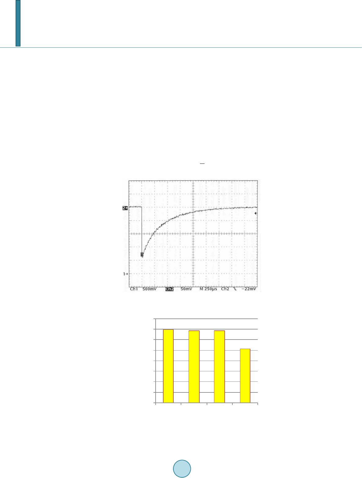 A. A. Komarskiy et al. commonly used to absorbed radiation has coefficient of X-ray radiation 0.01, as for intensifying screens Gd2O2S:Tb the coefficient is 0.29 [2]. Luminophors, which are used in the intensifying screens, have a certain period of persistence [3]. To define the duration of persistence precisely timing data and fluorescence intensity of luminophors, used in diagnostics, Kodak lanex, Renex EU-G3, Renex EU-G300, Renex EU-I4, were measured. Measuring was per- formed at a pulsed cathodoluminescence spectrometer. In Figure 1, you can see an oscillogram, picturing the process of luminophor persistence Renex EU-G3, typical for all the group of intensifying screens under investi- gation. The persistence duration of level 2.7 is 500 µs when pulse duration is 0.1 µs. The final results as persistence duration of the intensifying screens of level 0.1 are given in Figure 2. Measuring amplitude-time characteristics of persistence show, that all luminophors under investigation are characterized by the persistence at level of a millisecond, which is by an order of magnitude greater than radia- tion pulse duration of a nanosecond X-ray unit, as shown in Figure 3. X-ray quanta irradiate luminophor the X-ray photo detector luminophor with the intensity Ir, and the lumino- phor radiation intensity is (1) Figure 1. The persistence X-ray intensifying screen Renex EU-G3. Figure 2. Persistence duration of the intensifying screens of level 0,1. 0 200 400 600 800 1000 1200 1400 1600 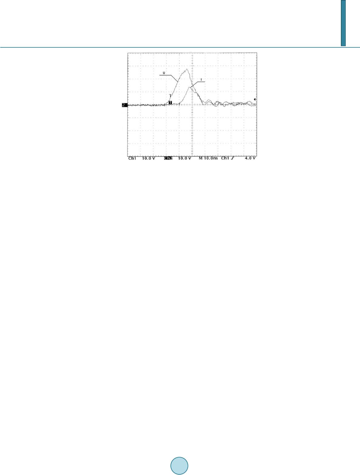 A. A. Komarskiy et al. Figure 3. An oscillogram of voltage and current of the X-ray tube RIA1-120. Voltage amplitude—110 kV, current ampli- tude—450 А, scale 10 ns/ point, repetition rate 850 Hz. where Il(t) is the radiation intensity at the time t, τ is the average time during which the excited state of lumino- phor atoms or molecules persists, T is period, I0 is radiation intensity at the time when excitation of lumines- cence stops (2) when using a constant X-radiation source, luminophor generates light radiation with the intensity I0 and a per- sistence fall after X-ray exposure in е times within (1 − 1.5)*10−3 (s) during the whole time of exposure from hundreds of milliseconds and higher. At pulse radiation after X-ray pulse exposure luminophor persistence cor- responds to е times during the same time, but this process repeats after each pulse. In the result of X-ray film accumulating light sum of the X-radiation, converted by luminophor, after X-ray pulse stops, the great differ- ence in duration of the X-ray pulse and the luminophor persistence fall makes it possible to reduce the exposure dose. At the Institute of Electrophysics of the Ural Division of the Russian Academy of Science we invented the pulse X-ray diagnostic portable apparatus 01 (XDPA 01) [4], which was based on the described effect. Then we upgraded the X-ray apparatus and created the pulse X-ray diagnostic portable apparatus Yasen-01 [5] [6]. We performed dosimetric tests, comparing the pulse X-ray diagnostic portable apparatus Yasen-01 (maximum voltage is 110 kV, the tube’s current is 150 A, pulse duration is 30 ns, repetition rate is 5 kHz) with the X-ray diagnostic apparatus Siemens Axiom Iconos R200 (maximum voltage is 125 kV), using a direct current tube. Measuring dosimetric parameters was carried out with the help of the dosimeter PTW UNIDOS 10001 with 30 cm3 cylindrical ionization chamber of 23361 kinds. Several standard modes of the tested machines for medi- cal diagnostics of different body organs were chosen. The main criterion for the comparison was the diagnostic quality of the X-ray photographs, as shown in Figure 4. Water phantom was used behind the ionization chamber to imitate a human body (as a scattered radiation source). Phantom thickness was 20 cm, focal distance to the ionization chamber was 80 cm. The results of in- vestigations are shown in Table 1. Then we performed tests, comparing the pulse X-ray apparatus Yasen-01 with the X-ray portable diagnostic apparatus Definium AMX 700 (maximum voltage is 105 kV) and with stationary X-ray complex Evolution HV which was created by STEPHANIX (maximum voltage is 40 - 150 kV). We compared the following parameters: effective doses, the quality of the X-ray images and usability. Calculating the effective doses of patients was made using the results of measuring radiation output of the apparatus with the help of the following measuring devices: the wide-range device for dosimetry of continuous and pulse X-rays and gamma rays DKS-AT1123, the all-mode dosimeter for controlling characteristics of X-ray apparatus-Unfors Xi RF&MAM detector. In Table 2, effective doses of patient irradiation are presented. Three proficient roentgenologists carried out independent estimation of the X-ray images’ quality and the ap- paratus’ usability. 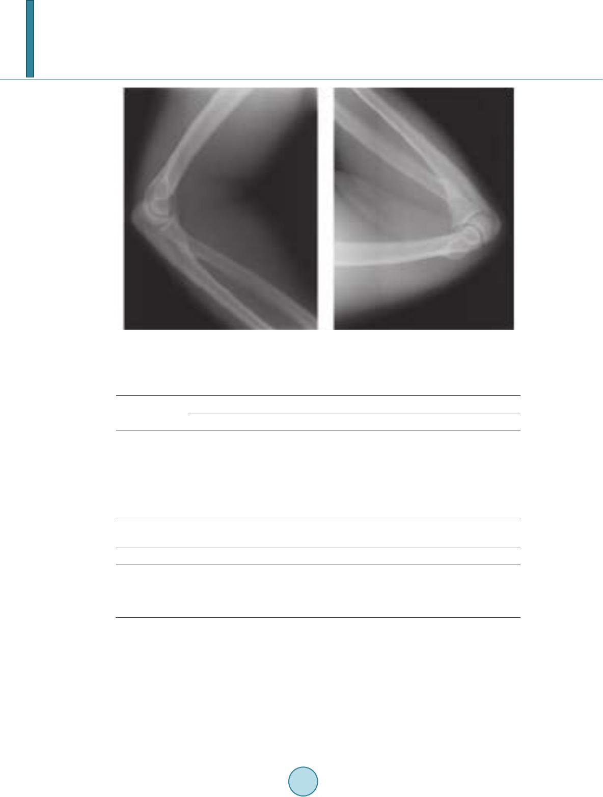 A. A. Komarskiy et al. (a) (b) Figure 3. Elbow arthrogram using by a) X-ray diagnostic apparatus with direct current tube; b) pulse X-ray diagnostic portable apparatus Yasen-01. Table 1. Test results of Siemens Axiom Iconos R200 and Yasen-01. Object, projection Siemens Axiom Iconos R200 Yasen-01 Voltage, kV Exposition, mAs Dose, µGy Voltage, kV Exposition, mAs Dose, µGy Lungs, direct 57 3.6 200 110 1.4 70 Skull, side 60 8 505 110 3.7 185 Thigh, direct 55 8 410 110 3.4 170 Knee, direct 47 6.3 215 100 1.8 86 Ankle, direct 48 4.5 160 100 1.0 48 Table 2. Effective doses of patient irradiation. Name Projection Effective dose, mSv Yasen-01 Chest (direct) 0.027 Definium AMX 700 Chest (direct) 0.570 Evolution HV Chest (direct) 0.721 3. Discussion As follows from the Table 1, the radiation dose of Yasen-01 is 2.5 - 3 times lower than the one of Siemens Axiom Iconos R200 and 20 - 25 times lower compared with Definium AMX 700, Evolution HV. The fact is that with the help of the pulse X-ray we can decrease the dose more significantly can be explained by the higher value of anode current (Yasen-01 – 150 A), since luminophor radiation intensity Il is proportional to X-ray quantum intensity Ir hence it proportional to the X-ray tube current Ie. Moreover, decreasing dose is explained by pulse duration: the Yasen-01 apparatus generates X-ray pulses of shorter duration (30 ns). Therefore the Ya- sen-01 apparatus has a more homogeneous radiation spectrum and greater effective energy (42.5 keV), i.e. it  A. A. Komarskiy et al. creates harder radiation. The X-ray tube’s performance in good conditions in terms of maximum acceptable heat load of the anode should be considered as one of the machines’ advantages. At present the operating mode of constant radiation machines is limited to 0.3 - 0.01 A. This limitation is subject to maximum acceptable heat load of the anode, since exceeding this value causes anode destruction. However the pulse tube current reaches the peak value of 150 А, which increases luminescence intensity Il by dozens of times. For example, if repetition rate is f = 1 kHz and pulse durations is τ = 10−8 s, duty factor (3) equals Q = 105, and the tube average current < I > (when current pulse is Ie = 100 А) is inversely proportional to pulse duty factor, ( 4) and equals just < I > = 1 × 10⁻³А. The chest X-Ray direct projection images, which were obtained at the pulse X-ray diagnostic portable appa- ratus Yasen-01, are the same as to the quality of the images, which were obtained by the X-ray portable diag- nostic apparatus Definium AMX 700 and are insignificantly inferior compared with the images obtained by the stationary X-ray complex Evolution HV which was created by STEPHANIX. Roentgenologist point out that thanks to the compact size and low weight (about 45 kg) the Yasen-01 appara- tus can be used in confined space and is easily transported. 4. Conclusions In conclusion, the conducted investigation demonstrates that X-ray radiation dose is decreased by several times thanks to using pulsed nanosecond X-ray machines as opposed to using relative conventional X-ray units. The dose of radiation was reduced so significantly thanks to the fact that persistence duration of X-ray radiation converters (luminophor) is several times higher than X-ray pulse duration. Further decreasing radiation dose is possible owing to increasing current pulse amplitude Ie, shortening pulse duration τ and increasing duty factor Q. The quality of the X-Ray images which were obtained at the pulse X-ray diagnostic portable apparatus Ya- sen-01 is the same as at constant X-radiation apparatus. Thanks to its small size and weight Yasen-01 apparatus can be used in confined space and easily transported in narrow corridors and elevators. References [1] Doroshko, M. V. (1998) New Capabilities in Reducing of Radiation Dose лу чев ой нагрузки. News of Radiodiagnos- tics, 3, 13-15. [2] Blinov, N.N., Bikov, R.E. and Kozlovskiy, E.B. (1989) Engineering Tools of Medical Introscopy. Leonov B.I., Moskow. [3] Kazgikin, O.N., Markovskiy, L.Y., Mironov, I.A., Pskerman, F.M. and Petoshina, L.N. (1975) Inorganic Phosphor. Leningrad. [4] Filatov, A.L., Korzhenevskiy, S.R., Kuznetsov, V.L., Ananin, M.V. and Motovilov, V.A. (2004) The Nanosecond PP X-ray Apparatus. Procedings of the 15th International Conference on High-Power Particle Beams, Saint-Petersburg, 1-7 April 2004, 552-554. [5] Filatov, A.L., Bastrikov, V.L., Korzhenevskiy, S.R., Kuznetsov, V.L. and Ponikaravskih, A.E. (2007) Universal Porta- ble X-Ray Apparatus. Russian Federation Patent No. 2005503167. [6] Mozharova I.E. and Kuznetsov V.L. (2011) The Pulse X-Ray Diagnostic Portable Apparatus “Yasen 01”. Medical business, 9, 84-85.
|