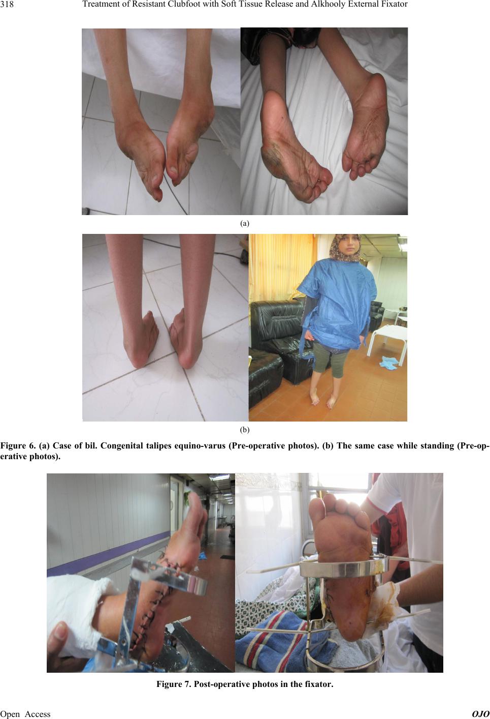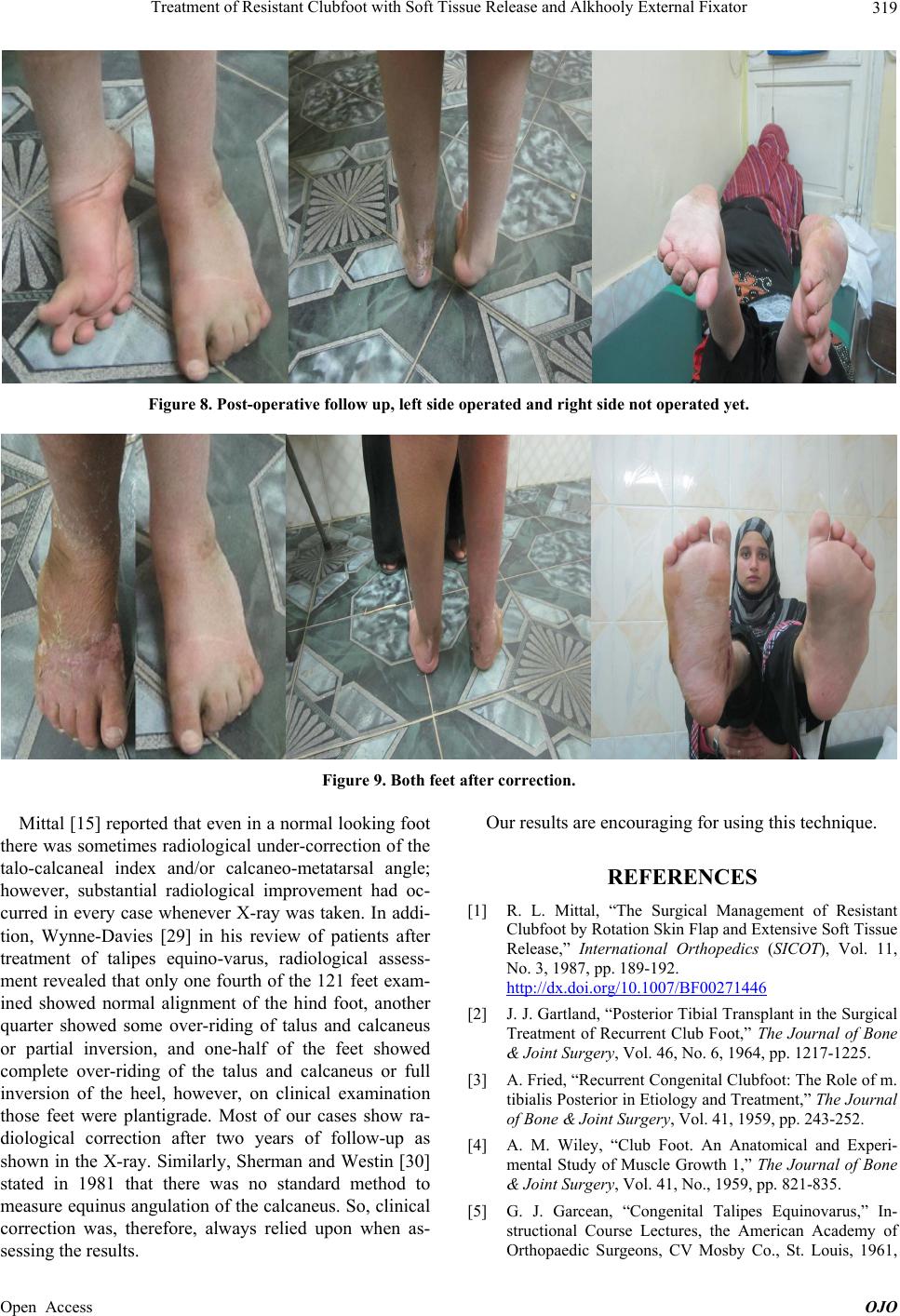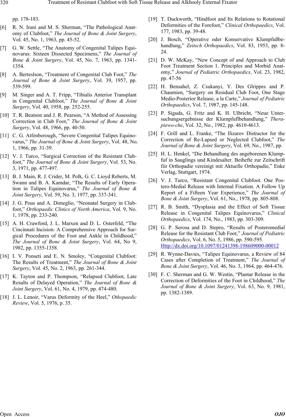
Treatment of Resistant Clubfoot with Soft Tissue Release and Alkhooly External Fixator
320
pp. 178-183.
[6] R. N. Irani and M. S. Sherman, “The Pathological Anat-
omy of Clubfoot,” The Journal of Bone & Joint Surgery,
Vol. 45, No. 1, 1963, pp. 45-52.
[7] G. W. Settle, “The Anatomy of Congenital Talipes Equi-
novarus: Sixteen Dissected Specimens,” The Journal of
Bone & Joint Surgery, Vol. 45, No. 7, 1963, pp. 1341-
1354.
[8] A. Berteslson, “Treatment of Congenital Club Foot,” The
Journal of Bone & Joint Surgery, Vol. 39, 1957, pp.
539-599.
[9] M. Singer and A. T. Fripp, “Tibialis Anterior Transplant
in Congenital Clubfoot,” The Journal of Bone & Joint
Surgery, Vol. 40, 1958, pp. 252-255.
[10] T. R. Beatson and J. R. Pearson, “A Method of Assessing
Correction in Club Foot,” The Journal of Bone & Joint
Surgery, Vol. 48, 1966, pp. 40-50.
[11] C. G. Atfenborough, “Severe Congenital Talipes Equino-
varus,” The Journal of Bone & Joint Surgery, Vol. 48, No.
1, 1966, pp. 31-39.
[12] V. J. Turco, “Surgical Correction of the Resistant Club-
foot,” The Journal of Bone & Joint Surgery, Vol. 53, No.
3, 1971, pp. 477-497.
[13] B. J. Main, R. J. Crider, M. Polk, G. C. Lioy d Re berts, M.
Swann and B. A. Kamdar, “The Results of Early Opera-
tion in Talipes Equinovarus,” The Journal of Bone &
Joint Surgery, Vol. 59, No. 3, 1977, pp. 337-341.
[14] J. G. Pous and A. Dimeglio, “Neonatal Surgery in Club-
foot,” Orthopaedic Clinics of North America, Vol. 9, No.
1, 1978, pp. 233-240.
[15] A. H. Crawford, J. L. Marxen and D. L. Osterfeld, “The
Cincinnati Incision: A Comprehensive Approach for Sur-
gical Procedures of the Foot and Ankle in Childhood,”
The Journal of Bone & Joint Surgery, Vol. 64, No 9,
1982, pp. 1355-1358.
[16] I. V. Ponseti and E. N. Smoley, “Congenital Clubfoot:
The Results of Treatment,” The Journal of Bone & Joint
Surgery, Vol. 45, No. 2, 1963, pp. 261-344.
[17] K. Tayton and P. Thompson, “Relapsed Clubfoot, Late
Results of Delayed Operation,” The Journal of Bone &
Joint Surgery, Vol. 61, No. 4, 1979, pp. 474-480.
[18] J. L. Lenoir, “Varus Deformity of the Heel,” Othopaedic
Review, Vol. 5, 1976, p. 35.
[19] T. Duckworth, “Hindfoot and Its Relations to Rotational
Deformities of the Forefoot,” Clinical Orthopaedics, Vol.
177, 1983, pp. 39-48.
[20] J. Bosch, “Operative oder Konservative Klumpfußbe-
handlung,” Zeitsch Orthopaedics, Vol. 83, 1953, pp. 8-
24.
[21] D. W. McKay, “New Concept of and Approach to Club
Foot Treatment Section 1. Principles and Morbid Anat-
omy,” Journal of Pediatric Orthopaedics, Vol. 23, 1982,
pp. 47-56
[22] H. Bensahel, Z. Csukanyi, Y. Des Glrippes and P.
Chaumien, “Surgery on Residual Club Foot, One Stage
Medio-Posterior Release, a la Carte,” Journal of Pediatric
Orthopaedics, Vol. 7, 1987, pp. 145-148.
[23] P. Siguda, G. Fritz and K. H. Ulbricht, “Neue Unter-
suchungsergebnisse der Klurnpfuflbehandlung,” Thera-
piewo-che, Vol. 32, No., 1982, pp. 4610-4613.
[24] F. Grill and L. Franke, “The Ilizarov Distractor for the
Correction of Re-Lapsed or Neglected Clubfoot,” The
Journal of Bone & Joint Surgery, Vol. 69, No., 1987, pp.
[25] H. L. Henkel, “Die Behandlung des angeborenen Klump-
fuf in Sauglings und Kindesalter. Beihefte zur Zeitschrift
fiir Orthopadie vereinigt mit Aktuelle Orthopadie,” Enke
Verlag, Stuttgart, 1974.
[26] V. J. Turco, “Resistant Congenital Clubfoot. One Pos-
tero-Medial Release with Internal Fixation. A Follow Up
Report of a Fifteen Year Experience,” The Journal of
Bone & Joint Surgery, Vol. 61, No., 1978, pp. 805-808.
[27] R. B. Smith, “Dysplasia and the Effect of Soft Tissue
Release in Congenital Talipes Equinovarus,” Clinical
Orthopaedics, Vol. 174, No., 1983, pp. 303-309.
[28] G. P. Serosa and D. Stepro, “Results of Posteromedial
Release for the Resistant Club Foot,” Journal of Pediatric
Orthopaedics, Vol. 6, No. 5, 1986, pp. 590-595.
Http://dx.doi.org/10.1097/01241398-198609000-00012
[29] R. Wynne-Davies, “Talipes Equinovarus, a Review of 84
Cases after Completion of Treatment,” The Journal of
Bone & Joint Surgery, Vol. 46, No. 3, 1964, pp. 464-476.
[30] F. C. Sherman and G. W. Westin, “Plantar Release in the
Correction of Deformities of the Foot in Childhood,” The
Journal of Bone & Joint Surgery, Vol. 63, No. 9, 1981,
pp. 1382-1389.
Open Access OJO