S. R. El-Shaboury et al. / Natural Science 2 (2010) 432-443
Copyright © 2010 SciRes. OPEN ACCESS
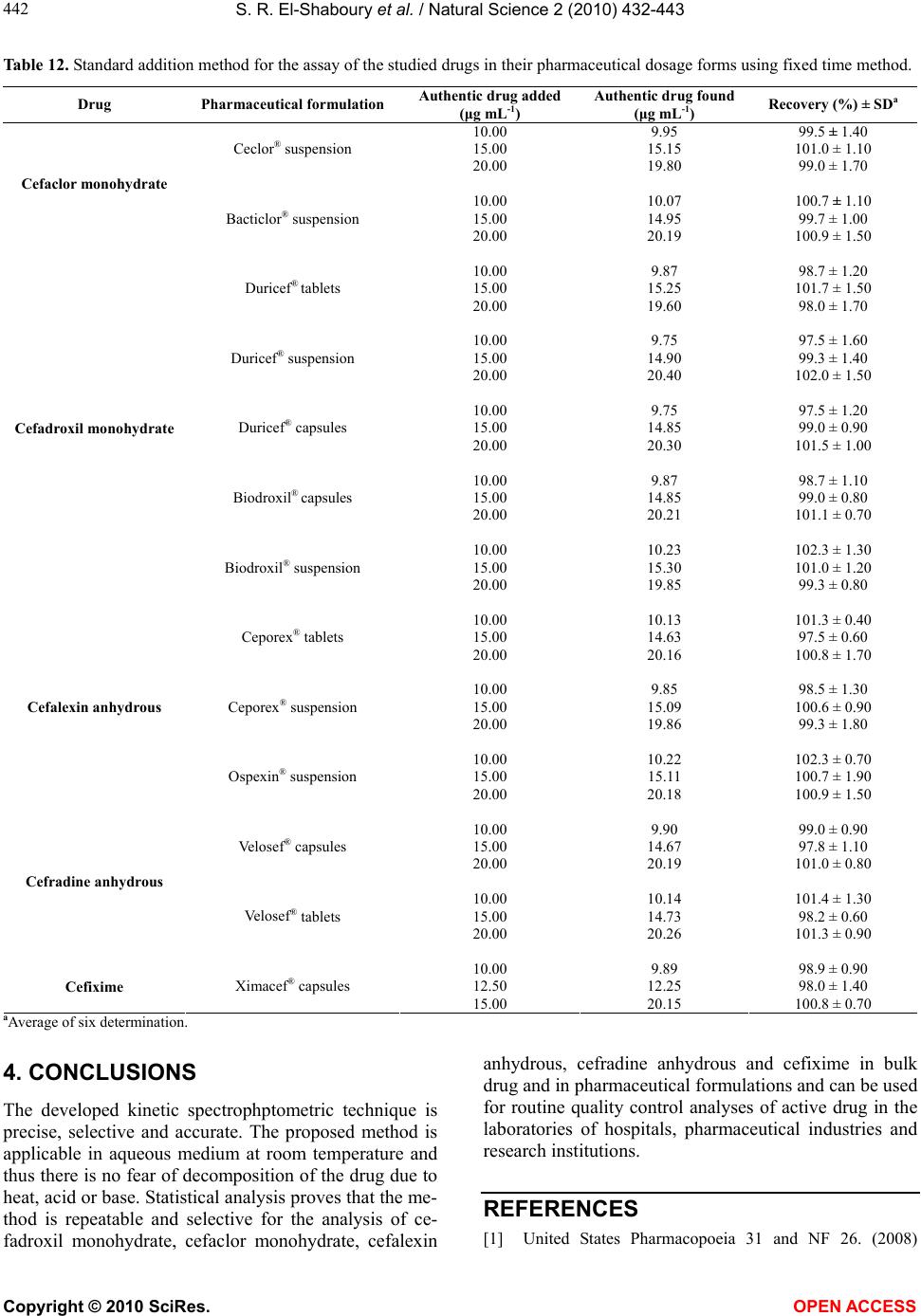
443
443
American Pharmaceutical Association, Washington, DC.
[2] El-Obeid, H.A., Gad-Kariem, E.A., Al-Rashood, K.A.,
Al-Khames, H.A., El-Shafie, F.S. and Bawaseer, G.A.M.
(1999) A selective colorimetric method for the determi-
nation of penicillins and cephalosporins with α-aminoa-
cyl functions. Analytical Letters, 32(14), 2809-2823.
[3] Metwally, F.H., Alwarthan, A.A. and Al-Tamimi, S.A.
(2001) Flow-injection spectrophotometric determination
of certain cephalosporins based on the formation of dyes.
Il Farmaco, 56(8), 601-607.
[4] Sastry, C.S.P., Rao, S.G., Naidu, P.Y. and Srinivas, K.R.
(1998) New spectrophotometric method for the determi-
nation of some drugs with iodine and wool fast blue BL.
Talanta, 45(6), 1227-1234.
[5] Ivama, V.M., Rodrigues, L.N.C, Guaratini, C.C.I and
Zanoni, M.V.B. (1999) Spectrophotometric determination
of cefaclor in pharmaceutical preparations. Quimica
Nova, 22(2), 201-204.
[6] Al-Momani, I.F. (2004) Flow-injection spectrophotomet-
ric determination of amoxycillin, cefalexin, ampicillin,
and cefradine in pharmaceutical formulations. Analytical
Letters, 37(10), 2099-2110.
[7] Yang, J., Zhou, G.J., Cao, X.H., Ma, Q.L. and Dong, J.
(1998) Study on the fluorescence characteristics of alka-
line degradation of cefadroxil, cefradine, cefotaxime so-
dium and amoxycillin. Analytical Letters, 31, 1047-1060.
[8] Aly, F.A., Hefnawy, M.M. and Belal, F. (1996) A selec-
tive spectrofluorimetric method for the determination of
some α-aminocephalosporins in formulations and bio-
logical fluids. Analytical Letters, 29(1), 117-130.
[9] Yang, J.H., Zhou, G.J., Jie, N.Q., Han, R.J., Lin, C.G. and
Hu, J.T. (1996) Simultaneous determination of cefalexin
and cefadroxil by using the coupling technique of syn-
chronous fluorimetry and h-point standard additions
method. Analytica Chimica Acta, 325(3), 195-200.
[10] Yang, J.H., Ma, Q.L., Wu, X., Sun, L.M., Cao, X.H. (1999)
A new luminescence spectrometry for the determination of
some β-lactamic antibiotics. Analytical Letters, 32(3),
471-480.
[11] Chailapakul, O., Aksharanandana, P., Frelink, T., Einaga,
Y. and Fujishima, A. (2001) The electrooxidation of sul-
fur-containing compounds at boron-doped diamond elec-
trode. Sensors and Actuators B, 80(3), 193-201.
[12] Chailapakul, O., Fujishima, A., Tipthara, P. and Siri-
wongchai, H. (2001) Electroanalysis of glutathione and
cefalexin using the boron-doped diamond thin-film elec-
trode applied to flow-injection analysis. Analytical Sci-
ences, 17(ICAS2001), i417-i422.
[13] Li, Q.L. and Chen, S. (1993) Studies on electrochemical
behaviour of cefalexin. Analytica Chimica Acta, 282(1),
145-152.
[14] Crouch, S.R., Cullen, T.F., Scheeline, A. and Kirkor, E.S.
(1998) Kinetic determinations and some kinetic aspects
of analytical chemistry. Analytical Chemistry, 70(12),
53R-106R.
[15] Perez-Bendito, D., Gomez-Hens, A. and Silva, M. (1996)
Advances in drug analysis by kinetic methods. Journal of
Pharmaceutical and Biomedical Analysis, 14(8-10),
917-930.
[16] Espinosa-Mansilla, A., Acedo Valenzuela, M.I., Salinas,
F. and Canada, F. (1998) Kinetic determination of
ansamicins in pharmaceutical formulations and human
urine; manual and semiautomatic (stopped-flow) proce-
dures. Analytica Chimica Acta, 376(3), 365-375.
[17] Helaleh, M.I.H. and Abu-Nameh, E.S.M. (1998) A ki-
netic approach for determination of cefadroxil in phar-
maceuticals by alkaline hydrolysis. Journal of AOAC In-
ternational, 81(3), 528-533.
[18] Rahman, N., Ahmad, Y. and Azmi, S.N.H. (2005) Kinetic
spectrophotometric method for the determination of
ramipril in pharmaceutical formulations. AAPS Pharm-
SciTech, 6(3), E543-E551.
[19] Neil, S.I. (1987) Physical organic chemistry. John Wiley
& Sons, New York, 93.
[20] Kelly, F.C. (1953) Studies on the stability of iodine
compounds in iodized salt. Bulletin of World Health Or-
ganization, 9(2), 217-230.
[21] Yatsimirskii, K.B. (1966) Kinetic methods of analysis.
Pergamon Press, London, 43.
[22] Laitinen H.A., Harris, W.E. (1975) Chemical analysis.
2nd Edition, McGraw-Hill, New York.
[23] (2005) Topic Q2 (R1): Validation of analytical proce-
dures: text and me thodology. International Conference
on Harmonization, Foster. http://www.ich.org/LOB/me-
dia/MEDIA417.pdf
[24] Saleh, G.A., Askal, H., Darwish, I. and El-Shorbagi, A.
(2003) Spectroscopic analytical study for the charge-
transfer complexation of certain cephalosporins with
chloranilic acid. Analytical Sciences, 19(2), 281-287.
[25] Harvey, D. (2000) Modern analytical chemistry. Boston,
McGraw-Hill, Massachusetts, 108.
[26] The Merck index (2001) An encyclopedia of chemicals,
drugs and pharmaceuticals. 13th Edition, Merck & Co.,
INC., New Jersey, 133.
[27] Svehla, G. (1979) Vogel’s textbook of macro and semi-
micro qualitative inorganic analysis. 5th Edition, the
Chaucer Press, Great Britain, 342.