Open Journal of Acoust i c s , 2011, 1, 55-62
doi:10.4236/oja.2011.13007 Published Online December 2011 (http://www.SciRP.org/journal/oja)
Copyright © 2011 SciRes. OJA
Ultrasound Por oelastic T issue Typing
Piero Chiarelli1,2, Bruna Vinci1, Antonio Lanatà2, Valeria Gismo ndi 2, Simone Chiarelli3
1National Council of Research of Italy, Pisa, Italy
2Interdepartmental Research Center E. Piaggio”, Faculty of Engineering,
University of Pisa, Pisa, Italy
3School of Engineering, University of Milan, Crema, Italy
E-mail: pchiare@ifc.cnr.it
Received October 4, 2011; revised November 12, 2011; accepted November 20, 2011
Abstract
Employing the poroelastic theory of acoustic waves in gels, the ultrasound (US) propagation in a gel medium
filled by poroelastic spherical cells is studied. The equation of fast compressional wave, the phase velocity
and the attenuation as a function of the elasticity, porosity and concentration of the cells into the gel matrix
are investigated. The outcomes of the theory agree with the preliminary measurements done on PVA gel
scaffolds inseminated by porcine liver cells at various concentrations. The feasibility of a non-invasive tech-
nique for the health assessment of soft biological tissues steaming by the model is analyzed.
Keywords: Ultrasound, Bi-Phasic Model of Living Tissues, Non-Invasive Ultrasound Assessment,
Poroelastic Ultrasounds in Soft Tissues
1. Introduction
The present work is motivated in obtaining a reliable
model for ultrasound (US) wave propagation in natural
soft tissues [1-4].
In preceding works the authors validated a poro-elastic
model for US propagation in synthetic as well as natural
gels such as those of the extra-cellular matrix of soft tis-
sues [4].
Even if the structure of a synthetic hydrogel is some-
how different from that of a soft biological mesh, there
exists a strong analogy between the macroscopic re-
sponse of charged hydrogels and that of living tissues,
such as for derma and cartilage [5], in the diffusional
wave limit.
On the base of this analogy, the US bi-phasic model for
soft tissues appears to be a promising tool for the deve-
lopment of non-invasive health assessment methods and
for the study and the characterization of tissue mimicking
phantom for US thermal therapy [6-8].
This fact is confirmed by the current research that
clearly shows how the knowledge of the link between the
poroelastic characteristics of a biological tissue and its
acoustical behavior is a source of information that can be
used for non-invasive investigations [8-10].
Nowadays, the US propagation in natural hydrogels,
mostly composed of water, is usually modeled by means
of the wave equation that holds for liquids [11].
This approach is poorly satisfying because it com-
pletely ignores the liquid-solid arrangement, and it does
not completely explain the experimental behavior of the
US propagation.
Moreover, there are many discrepancies between US
propagation in water and in soft tissues both for trans-
verse and longitudinal acoustic waves [1].
Actually, tissues are modeled as water solutions of
natural polymers and proteins that may have bounded
resonant states [1] with the US attenuation showing a fre-
quency (ν) law [12]: ν(1 +
), with ranging between 14
and 12 (
= 1 for water).
At high frequencies the classical poroelastic theories,
mainly developed for geological studies, [13-22] lead to
a dependence of attenuation with
= 1 without any pos-
sibility to have a fractional value of
.
The recent gel bi-phasic model for US [4] shows that
is possible to have a frequency dependence of attenua-
tion with fractional values of as a direct consequence
of the presence of bounded water onto the polymer net-
work that affects the friction behavior of the fluid against
the solid matrix.
At high frequencies the bounded water bearing lowers
the friction between the free water and the polymer ma-
trix leading to the peculiar behavior of the US phase ve-
locity and attenuation of gels.
P. CHIARELLI ET AL.
56
Actually, the natural tissues are far to be homogenous
but may present anisotropies, blood vessels and cells. In
the present work spherical cells are introduced into the
substrate of a homogeneous extra-cellular hydrogel ma-
trix. The biological cells are designed as poro-elastic
spheres, endowed by internal and superficial elasticity as
well as permeability, and are assumed to be isotropically
dispersed in the hydrogel environment. The model is de-
veloped in the continuum limit approach for US wave-
length much bigger than the cells dimension (typically
up to about 10 MHz).
The model presented here discloses how the behavior
of the US propagation is linked to the arrangement of the
biological medium and its poroelastic characteristics.
This papers shows that this fact can be used for the
development of non-invasive health assessment techni-
ques for tissues and organs by monitoring the elasticity,
porosity and water content of the cells and the extra-
cellular matrix. The detection of the liver cirrhosis is the
short term application proposed in this paper [6].
2. Poro-Elastic US Wave in Soft Living
Tissues
2.1. US Wave in Highly Hydrated Gels
The poroelastic wave equations for hydrogels can be ob-
tained by introducing the appropriate fluid network inte-
raction to take into account for the bounded water pre-
sence around the polymer chains [4].
Under the assumption that the bounded water volume
fraction is very small and that the “polymer-bounded
water aggregate” constitutes the solid matrix of the bi-
phasic mean, it is possible to obtain with the following
poroelastic motion equations [4]
 

()
e
f
ee te
e
  
 



  

t
(1a)


22
11 12
e
PQe et
2
f
ee t
 
 

 

(1b)


22
12 22
Re
e
Qe
2
t
f
ee t
 
 

 

(1c)
e

 (2)
where
ij is the solid strain tensor, eαα is the trace of the
liquid strain tensor, e
is the trace of the bounded wa-
ter strain tensor; P, Q, and R are the poroelastic medium
constants that can be measured by means of jacketed and
unjacketed experiments [18]; β is the water volume frac-
tion of the hydrogel,
is the volume fraction of bounded
water, f is the inverse of the hydraulic permeability of the
matrix [18], and
11,
12 and
22, are the mass densities
defined as:
11 + 2
12 +
22 =
,
11 +
12 = (1 – βe)
s,
12 +
22 = βe
f; where
s and
f represent the solid and
the liquid mass densities respectively, while
is the total
mass density of the biphasic medium. Moreover, the ela-
stic constant
and the friction coefficient
describe the
polymer-bounded water interaction.
The above equations are derived by assuming that [4]:
1) The inertial effect of bounded water can be disre-
garded;
2) The trace of the strain tensor of the polymer
approximates that one of the solid aggregate.
Moreover, by introducing the condition that the water
content in the hydrogel is very high, further approxima-
tions can be introduced into the wave equations such as
RQP
(3)
11
ee e
QR
 
(4)
1211 22

(6)
to obtain
222 2
22
11
(Re )()
()
eef
e
et
et
 
 


 

(6a)
22
(R)( )
ef
f
ee tQe t
 

  (6b)


()
e
f
ee te
e
  
 



t

 (6c)
For the fast plane wave ()
x
ikxt
Ce e

Equation
(1a) reads
() ()
e
f
ee tFe
  

t
 (7)
where the complex friction coefficient of the gel F()
reads

1
1
1
()()() j()
e
Ff
 

(8)
Leading, by Equation (6a), to the characteristic equa-
tion

22 3
11( )
() 1(1)
fe e
kiR iF
 
 (9)
that transformed In two real equations in
and c gives

22 2221
01
11(1)Im
ee
cc kF


 1()
(10)
 
21
011
21R
e
ak ccF

 
()
e (11)
where

032
f
c
c

(12)
is the pure elastic longitudinal US wave velocity in the
Copyright © 2011 SciRes. OJA
P. CHIARELLI ET AL.57
gel,

12
ff
cR
the US velocity in the intermolecu-
lar fluid (free water) and


2
1222
Re1 ee
Fff


 

(13)


2
12
Im()F
 
 (14)
Assuming that the polymer-bounded water viscosity
(
) follows the frequency behavior [4]
()0g

(15)
with 0 <
1/2 and where 0
2π
g
f

, it follows
that

g
1
1
lim Re1F(
)

(16)

g
1
1
lim Im0F

(17)
Thence, in a hydrogel with a infinitely dilute polymer
matrix (βe 1 and 11 1
) Equations (10) and (11) read

22 22
0
1cck c
 
2
0
(18)
where since typically (
/k)2 is very small (of or-
der of 10–3 in hydrogels), and
2
0
cc

 
21
0
21
p
f
kcc


  (19)
where
11
2π
p
ff

(20)
Finally, when the polymer network is not very diluted,
but has a real finite concentration, the series expansion as
a function of the fractional polymer volume (1 - β) can
be introduced into Equation (12) to read [4]

2
2
03
12
(1 )(1)
f
c
c2
 
 (21)
2.2. US Wave Equation in a Hydrogel with
Dispersed Cells
When we describe a tissue as a hydrogel biphasic mean
containing cells, we have to refer to the overall tissue con-
stants βt, Rt,
ft, Ft and so on, into the Equations (6a)-(6c)
that for plane waves lead to
222 13
0
() (1)
tt
cee tFet
 

 
3
11
(22)
where
3
0
tf
cR t

(23)
The constants βt Rt and
ft affect the phase velocity of
the elastic limit, while βt and Ft affect the US absorbance.
As far as it concerns βt, it influences the US attenuation
Berryma matrices [23] for which
11 (1 –
t)
s.
By defining
as the fractional volume of cells in
through
11 as it can be explicitly shown in diluted
n
side
th
me of cells/Total volume of tissue,
the m
e tissue to read
= Total volu
ean fractional water content of the tissue βt reads

(1)1- 1
tecece

  (24)
where βc is the fractional cells free water content.
.3. Us Speed in Natural Tissue
order to investigate the US phase velocity in the hy-
ompressibility modulus R
an
ng that the velocity cft = R/
ft slightly changes
as
2
In
drogel-cells syncytium we need to determine the biphasic
parameters concerning the inertial and the elastic terms
in the motion Equations (22).
As far as it concerns the ct
d the mass density
ft of the syncytium they are influ-
enced by the chemical (e.g., ionic strength) and mass
composition of the cells that usually differ from that ones
of the extra-cellular matrix. Thence, cft = (Rt/
ft)1/2 and
the elastic phase velocity ct0 are function of cell concen-
tration
.
Assumi t
a function of the cells concentration
, the series ex-
pansion

12 2
1
ftt ftf
cRc AA

 (25)
can be retained for the tissue.
uations (18) and (21) for a
bi
Moreover, analogously to Eq
ological tissue we can assume

12
2
1( )cc k3
00tttt ftt
c c

 (26)
and, hence,
23
1
tf
ccAAt

 (27)
where ct0 is the pure elastic phase velocity in the tissue
.4. Us Attenuation in Natural Tissue
this section we derive the complex friction coefficient
duc-
ta
and
t is the absorbing coefficient we are going to calcu-
late in the next paragraph.
2
In
F(
) (the inverse of hydraulic conductance of the syn-
cytium) that is responsible for the US attenuation.
We assume the following complex hydraulic con
nce:
1)

11
g
g
F
for the extra-cellular hydrogel;
1
2)

1
b
b
F
for the internal jelly cell body;
3)

1
m
F
for the cel
sinuy
/2 we assume
ls membrane.
Forsoidal inputs of frequenc
Copyright © 2011 SciRes. OJA
P. CHIARELLI ET AL.
58
that

11
mm
m
F
KibE

 (28)
where the real part Km is the hydraulic permeability of
inputs has
al
of cells of radius “a” embedded in
a
the cell membrane and where the imaginary one is its
superficial compliance proportional to the inverse of
Young’s elastic modulus Em. In the case where the cell
membrane thickness is much smaller than the cell dia-
meter, b–1 represents the membrane thickness.
Since the problem of sinusoidal electrical
ready been solved [24], we can easily find the overall
conductance of the tissue by making use of the electric-
hydraulic analogy.
Given a syncytium
hydrogel with the following electrical parameters
1)
g
electrical conductance of the extra-cellular
hydro
2) b
gel;
electrical conductance of the internal body of
the cell;
3) m
surface electrical conductance of the cells
m
rface electrical capacity of the cells mem-
br
global electrical admittance Y of the syncytium
[2
emb;
4) m
C su
rane
ane.
The
1] reads

2(1)(12)
(2)(1 )
g
b
g
g
Y
b
m
 

  (29)
where
()
bmb mbm
aY aY

 (30)
and where
m
C
mm
Yi
 (31)
Therefore by mean of the following substitutions
1) 1
g
g
F
hydraulic admittance of the extra-cellu-
lar hy
2) b
drogel;
1
b
F
of the cell;
hydraulic admittance of the internal
body
3) mm
K
surface hydraulic admittance of the
cells m
surface elasticity of the cells mem-
br
nd up with the global hydraulic admittance of ce-
llu
embrane;
4) 1
mm
CbE
ane.
We e
lar syncytium

11
11
1
2(1)(12)
(2)(1)
gb
g
gb
FF
FF FF





 
1
m
(32)
where 1
bm
F
ds
is a combined bulk-membrane permeability
that rea

1111
bm bmbm
1
F
FbaFF aF
 
 (33)
In order to apply the above model to a bio
su
l membrane of the cells has a very
lo
logical tis-
e we need to single out the relative magnitude of the
hydraulic constants.
Since the superficia
w hydraulic permeability (it separates the inner cell
body from the external hydrogel matrix), we expect that
the frequency
m/2 is not very high.
Therefore, at high frequencies 1
m
mKmbE

,
th e prevails on its per
1
m
e compliance of the cell membran -
meability and it follows that
11
mmm
F
KibE ibE


 (34)
that

11 11
1
bm bbm
F
FFi baE
 
 (35)
and that the hydraulic admittance of the tissue reads


11
2(1)(12)
 


11
11
11
(2 )(1)1
gb
g
gbbm
F
aib E
  




 
(36)
If we assume 1
b
F
to r
to have the typical form of E
tio
qua-
ns (15) and (16)ead

() 0bb bgb
F


 , (37)
with 0
2π
g
bbfb

, it follows that


11 1 1 11
11
bmbbmbbgb
FaibE i

  
 
 
(38)
where
0bm
Eab
b

(39)
Since
< 1, at very high frequencies,



11
bbg

the imaginary part of Fbm tends to vanis, so that h
bm b
F
(40)
and hence

11
11
1
2(1)(12)
(2)(1 )
gb
g
g
F
 




 
1
b
(41)
The friction coefficient of the syncytium given by the
expression (41) (as a whole) leads to the US attenuation
that reads

21
)11
2(1)(1)
ttft e
kcc F

 Re (42)
where
represents the normalized difference of water
content between the cells and the extra-cellular matrix
that reads
 
1
ec e
 
  (43)
It is useful to note that for βe close to 1, can be nega-
Copyright © 2011 SciRes. OJA
P. CHIARELLI ET AL.59
tiv
change of the normalized
U
g
e and even bigger than one.
In Figure 1 it is shown the
S attenuation as a function of the fraction of the cell
volume
for two values of the permeability ratio be-
tween cell body and the extra-cellular matrix:
b/
g =
0.01 and
b/
g = 0.1.
In the case b

, the overall friction coefficient
re
ads

11
12 1
g
F





(44)
while for bg

reads

11
1
12
g
F




(45)
When the cellular volume is a small part
vo
of the total
lume of the tissue (1
) it follows that

111 1
12 32F
 
 
 
1 1
ggbgb
 
 
(46)
That for bg

leads to
13
g
F11


, (47)
while for bg

gives
1
F

1
13 2
g

(48)
In Figure 2 it is shown the change of
U
the normalized
S attenuation as a function of the fraction of the cell
volume
for three values of the permeability ratio:
100
bg

, 10
bg

2
bg

.
3 it tIn Figureis shownhe US attenuation for a value
of
= 0.80 typical of the liver tissue as a function of the
permeability ratio bg

varying from 10–2 to 100.
By comparing fo(47) with (48) we can see thrmula at a
very different behavior for the US attenuation happens
whether or not b
is bigger than
g
. The angular coe-
fficient of the 48) changes from
positive (+3) for bg
-linear relations (47,

, to negative (–3/2) for
bg

3. Experimental
.1. Materials and Methods
el samples were prepared dissolving 0.5 ml of an aque-
0˚C for 24 h
an
va
3
G
ous solution of sodium alginate at a concentration of 2%
by weight (Alginic acid sodium salt from brown algae,
Sigma A0682-1006) in 0.5 ml of CaCl2 solution (FLU-
KA 06991) at a concentration of 0.4% by weight to ob-
tain the cross-liking of the polymer matrix.
The gel samples were refrigerated at –2
d then lyophilized at –40˚C under vacuum for 12 hours.
The gel samples were inseminated by liver cells at
rious densities: 105 cells/cm3, 2 × 10⁵ cells/cm3, 5 × 10⁵
cells/cm3, 106 cells/cm3, 2 × 10⁶ cells/cm3, 5 × 10⁶ cells/cm3,
Figure 1. Change of the normalized US attenuation as a
function of the fraction of the cell volume
for two values of
the permeability ratio:
b/
g = 0.01 and
b/
g = 0.1.
Figure 2. Change of the normalized US attenuation as a
function of the fraction of the cell volume
for the values of
the permeability ratio:
b/
g = 100,
b/
g = 10,
b/
g = 2.
Figure 3. US attenuation for
= 0.80 as a function of the
permeability ratio
b/
g varying from 10–2 to 100.
Copyright © 2011 SciRes. OJA
P. CHIARELLI ET AL.
60
then in a
lses were generated by the Panamet-
ric
in the ex-
pe
nal registration and conditioning data were co-
lle
amples were totally dehydrated in an oven
at
duced by
using the m
placed into an incubator at for 30 minutes and
refrigerator at 4˚C.
The ultrasonic pu
s® Pulser model 5052PR coupled with a PVDF pie-
zoelectric transducer obtained in our laboratory follow-
ing the Naganishi e Ohigashi procedure [25].
The frequencies of the ultrasonic wave used
rimental test were of 1.4 MHZ and 1 MHz. The dis-
tance between the transducer and the reflecting iron layer
behind the samples was measured with an accuracy of
0.01 cm.
Echo Sig
cted with a routine and carried out with the LabView™
software on a computer through a National Instruments®
DAQ device.
Finally, the s
40˚C with desiccant silica gels, to measure their poly-
mer content and the US speed in the dry solid.
The US absorption coefficient “
” was de
athematical relation 0
(2 )
1ln
2d
A
dA
A0 and A(2d) represent both the initial and final wave am-
, where
plitude, respectively, and where d is the sample thickness.
The water volume fraction of the hydrogel samples

w
VVV, where w
V and
w p
p
V are the volume
lymer respctivelywas obtained by
means of the respective weight fractions w
P and
of water and poe,
p
P
such as

w
PPP
 since the waternd PV
specific deose each other.
The fitting of the experimental results wer
w
nsities are very
e carried out
by
.2. Measurement of Ultrasound Wave
troducing the measured values βe = 0.9 for our extra-
is value in Equation (42), the best-
fit
p
cl
aA
means of a multiple parameter best fit utilizing an
appropriate routine in MATLAB®7.0.
3Attenuation
In
cellular PVA scaffold and βc = 0.89 for the cells in (43),
we obtain
0.1.
By introducing th
ted curve of experimental US attenuation in Figure 4
has been obtained for the ratio 6.54
bg

.
The result put in evidence tpehat once the rmeability
of the extra-cellular gel scaffold 1
g
is known or mea-
sured, the US poro-elastic model as deriving the per-
meability of the cellular bulk 1
b
llow
.
4. Discussion
he acoustic bi-phasic model for soft living tissues de-
T
scribes the US propagation in terms of collective cells
and extra-cellular matrix characteristics such as: 1) The
permeability and the elasticity of the cells and of the extra-
Figure 4. Experimental values of the normalized US at-
ellular matrix; 2) The percentage of cellular volume of
ave speed, the model does
no
tenuation in PVA-porcine liver cells composites with the
best fit obtained for
b/
g = 6.54 as a function of the frac-
tion of the cell volume
.
c
the tissue; 3) The fractional volume of water of cells and
of the extra-cellular matrix.
As far as it concerns the w
t make an explicit derivation of the coefficients A1 and
A2 by the constituents of the cellular syncytium. On the
contrary, the model details how the US attenuation de-
pends by the cell elasticity and permeability through the
term
1
Re F.
By using ts inhiformation, it is possible to define an
ex
r, since the poro-elastic characteristics of the
ce
pe of the absorbance frequency
sp
perimental method for the measurement of the perme-
ability of the cells once that one of the external scaffold
is known.
Moreove
lls and extra-cellular matrix may appreciably change in
pathological states (e.g. as in the cirrhosis) in principle,
the health state of biological tissues can be tracked by
means of ultrasounds.
In particular, the sha
ectrum determined by the characteristic frequencies
0
2π
g
bbfb

,
0
bm
Eab
b
and
1
mmm
K
Eb
can give information about the health state of cells and
. Concluding Remarks
he bi-phasic continuum model for US propagation in
tissues (as a sort of eco-biopsy) on the basis of epidemi-
ological comparisons.
5
T
hydrogels has been used to build up the acoustic wave
Copyright © 2011 SciRes. OJA
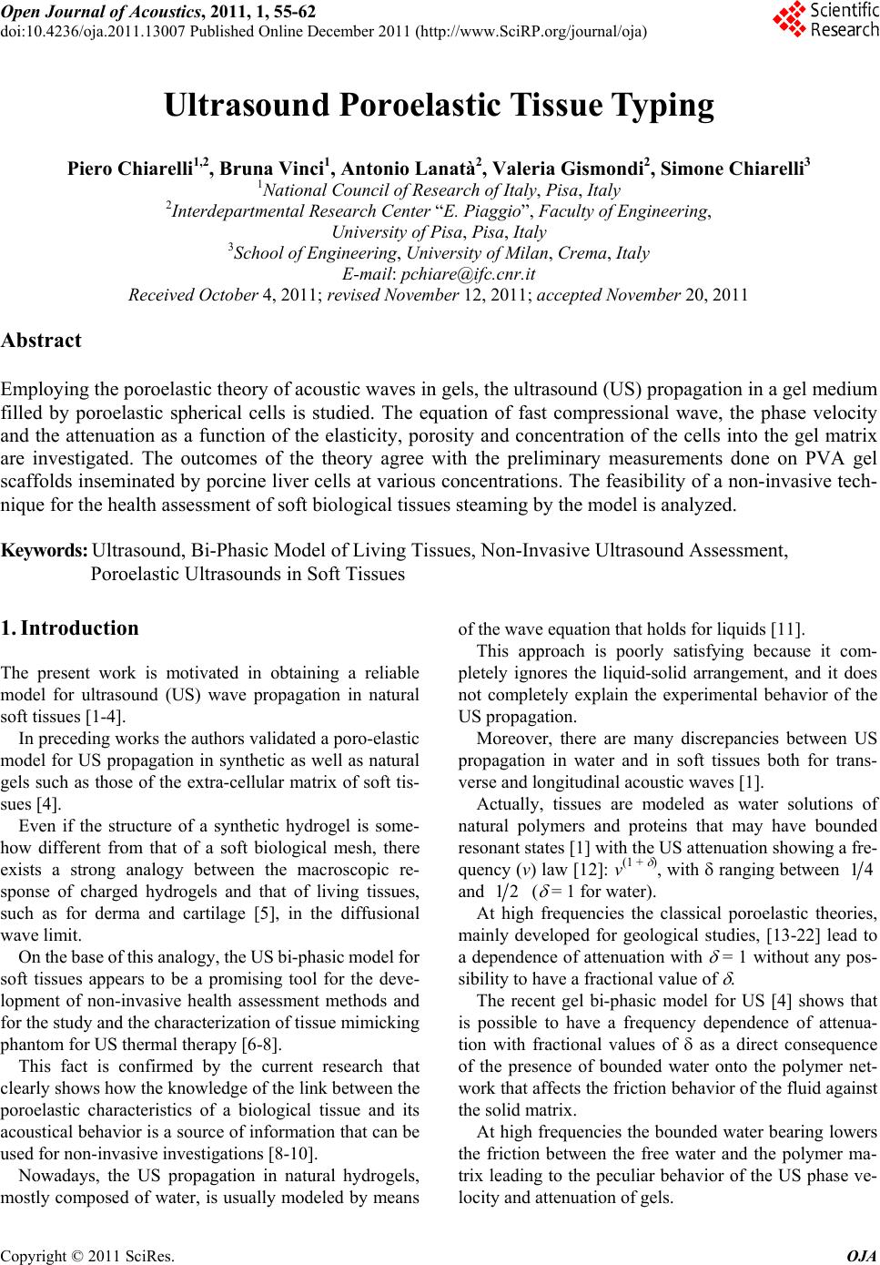
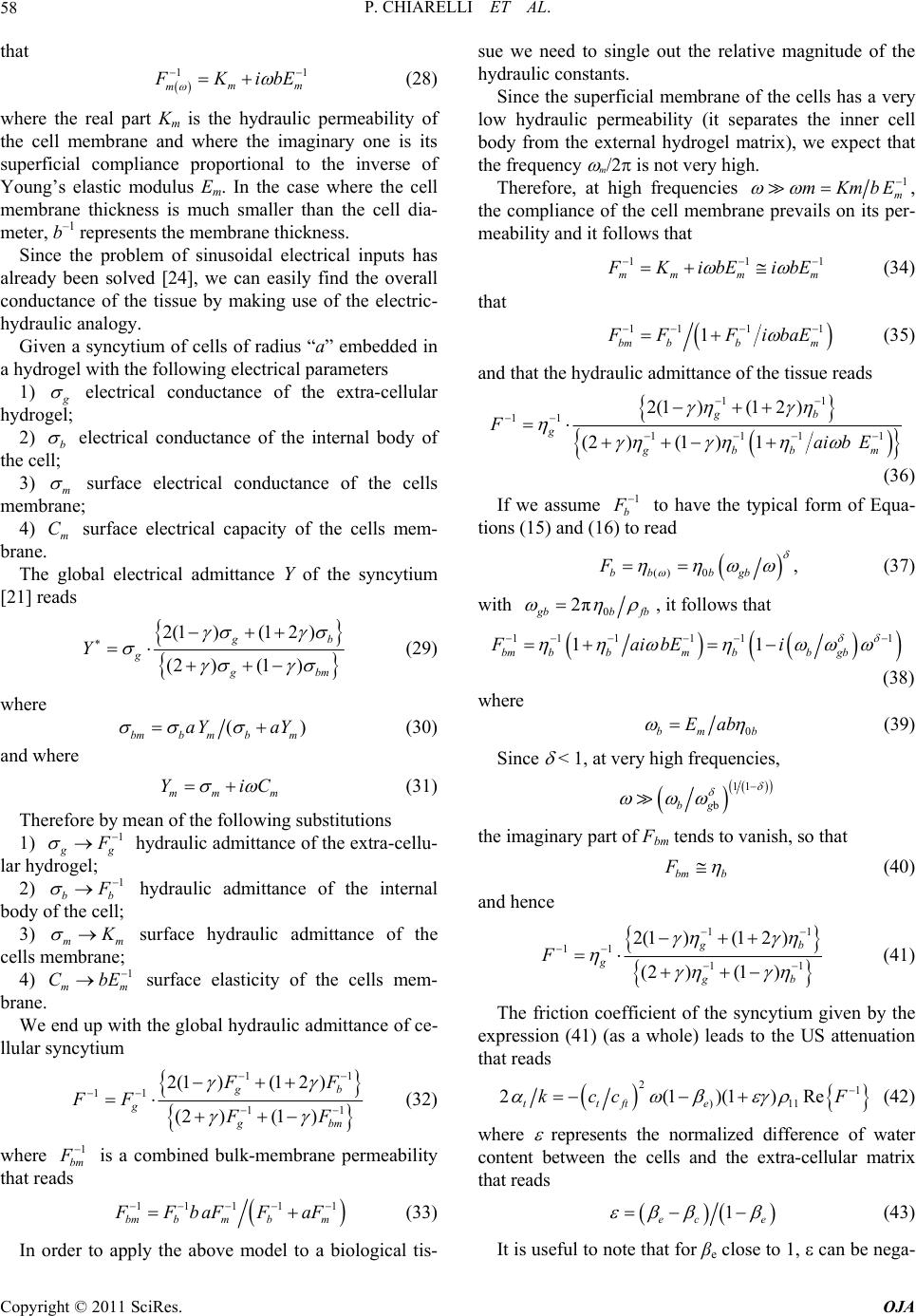
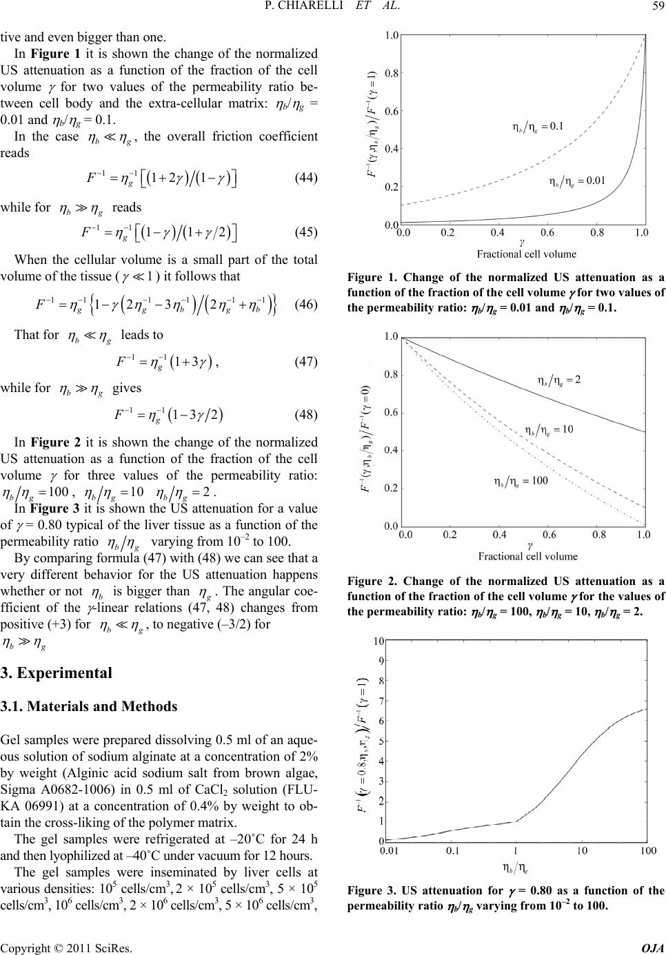
P. CHIARELLI ET AL.61
odel shows that the absorption of US is sensitive
to
model shows that the spectrum of US absorption
in
minary measurements
do
. References
] F. A. Duck, “Acoustic Properties of Tissue at Ultra
kau, R. W. Barnes and C. P. McGraw,
equation for a tissue-like syncytium made of spherical
cells homogeneously immersed in an extra-cellular gel
matrix.
The m
the cellular content of the tissue as well as of the po-
rosity of the cells body with respect to the external ma-
trix.
The
a biological tissue has a characteristic shape depend-
ing by the elasticity and permeability of cells and extra-
cellular matrix. By means of these parameters that are
linked to the health state of a tissue, the model can be
used to monitor pathologies of it.
The model agrees with preli
ne on porcine liver cells embedded in a poly-vinyl-
alcohol matrix. The experimental results have put in evi-
dence that the porcine liver cells have the bulk perme-
ability lower than that one of the PVA gel scaffold.
6
[1 sonic
Frequencies,” Academic Press, London, New York, 1990,
pp. 75-99.
[2] F. W. Krem “Ul-
trasonic Attenuation and Propagation Speed in Normal
Human Brain,” Journal of Acoustical Society of Ameri-
can, Vol. 70, No. 1, 1981, pp. 29-38.
doi:10.1121/1.386578
[3] J. W. Wladimiroff, I. L. Craft and D.G. Talbert, “In Vitro
Measurements of Sound Velocity in Human Fetal Brain
Tissue,” Ultrasound in Medicine & Biology, Vol. 1, No. 4,
1975, pp. 377-382. doi:10.1016/0301-5629(75)90125-8
[4] P. Chiarelli, et al., “High Frequency Poroelastic Waves in
Hydrogel,s” Journal of Acoustical Society of American,
Vol. 127, No. 3, 2010, pp. 1197-1207.
doi:10.1121/1.3293000
[5] D. De Rossi, A. Nannini and C. Domenici, “Artificial
Sensing Skin Mimicking Mechanoelectrical Conversion
Properties of Human Dermis,” IEEE Transaction on Bio-
medical Engineering, Vol. 35, No. 8, 1988, pp. 3-92.
doi:10.1109/10.1343
[6] S. Lochhead, D. Bradwell, R. Chopra and M. J. Bronskill,
er, K. Braun, T. Dreyer, P. Huber
al., “Noninvasive assessment of Liver Fibrosis
“A Gel Phantom for the Calibration of MR-Guided Ul-
trasound Thermal Therapy,” Proceedings of 2004 IEEE
Ultrasonics Symposium, Montreal, Vol. 2, 23-27 August
2004, pp. 1481-1483.
[7] G. Divkovic, M. Liebl
and J. Jenne, “Thermal Properties and Changes of Acous-
tic Parameters in an Egg White Phantom during Heating
and Coagulation by High Intensity Focused Ultrasound,”
Ultrasound in Medicine Biology, Vol. 33, No. 6, 2007, pp.
981-986.
[8] M. Ziol, et
by Measurement of Stiffness in Patient with Chronic He-
patitis C,” Hepatology, Vol. 41, No. 1, 2005, pp. 48-54.
doi:10.1002/hep.20506
[9] G. P. Berry, J. C. Bamber, C. G. Armstrong, N. R. Miller
edbio.2006.01.003
and P. E. Barbonne, “Toward an Acoustic Model-Based
Poroelasticity Imaging Method: I. Theoretical Founda-
tion,” Ultrasound in Medicine Biology, Vol. 32, No. 4,
2006, pp. 547-567.
doi:10.1016/j.ultrasm
upersonic Shear [10] J. Bercoff, M. Tanter and M. Fink, “S
Imaging: A New Technique for Soft Tissue Elasticity
Mapping,” IEEE Transactions on Ultrasonics, Ferroelec-
trics and Frequency Control, Vol. 51, No. 4, 2004, pp.
396-409. doi:10.1109/TUFFC.2004.1295425
[11] M. L. Mather and C. Baldock, “Ultrasound Tomography
Imaging of Radiation Dose Distributions in Polymer Gel
Dosimeters: Preliminary Study,” Medical Physics, Vol.
30, No. 8, 2003, pp. 2140-2148. doi:10.1118/1.1590751
[12] X. Yang and R. O. Cleveland, “Time Domain Simulation
M. Courdille, J. Dumas and R. Rajaonari-
roperties of Tissue at Ultrasonic
eneral Theory of Three-Dimensional Con-
of Nonlinear Acoustic Beams Generated by Rectangular
Piston with Application to Harmonic Imaging,” Journal
of Acoustical Society of American, Vol. 171, No. 1, 2005,
pp. 113-123.
[13] J. C. Bacri, J.
son, “Ultrasonic Waves: A Tool for Gelation Process
Measurements,” Journal of Physique Letters, Vol. 41, No.
15, 1980, pp. 369-372.
[14] F. A. Duck, “Acoustic P
Frequencies,” Academic Press, London, New York, 1990,
pp. 112-113.
[15] M. A. Biot, “G
solidation,” Journal of Applied Physics, Vol. 12, No. 2,
1941, pp. 155-164. doi:10.1063/1.1712886
[16] M. A. Biot, “Theory of Propagation of Elastic Waves in a
of Propagation of Elastic Waves in a
tic Coefficients of the Theory of
Gels,” Journal of Che-
Fluid-Saturated Porous Solid. II. High Frequency Range,”
Journal of Acoustical Society of American, Vol. 28, No. 2,
1956, pp. 179-191.
[17] M. A. Biot, “Theory
Fluid-Saturated Porous Solid. I. Low-Frequency Range,”
Journal of Acoustical Society of American, Vol. 28, No. 2,
1956, pp. 168-178.
[18] M. A. Biot, “The Elas
Consolidation,” Journal of Applied Mechanics, Vol. 24,
No. , 1957, pp. 594-601.
[19] D. L. Johnson, “Elastodynamics of
mical Physics, Vol. 77, No. 3, 1982, pp. 1531-1539.
doi:10.1063/1.443934
[20] R. N. Chandler, “Transient Streaming Potential Measure-
ments on Fluid-Saturated Porous Structures: An Experi-
mental Verification of Biot’s Slow Wave in the Quasi-
Static Limit,” Journal of Acoustical Society of America,
Vol. 70, No. 1, 1981, pp. 116-121. doi:10.1121/1.386689
[21] A. Peters and S. J. Candau, “Kinetics of Swelling of Sphe-
rical and Cylindrical Gels,” Macromolecules, Vol. 21, No.
7, 1988, pp. 2278-2282. doi:10.1021/ma00185a068
[22] D. L. Johnson, “Equivalence between Fourth Sound in Li-
quid He II at Low Temperature and the Biot Slow Wave
in Consolidated Porous Media,” Applied Physics Letters,
Vol. 37, No. 12, 1980, pp. 1065-1067.
Copyright © 2011 SciRes. OJA
P. CHIARELLI ET AL.
Copyright © 2011 SciRes. OJA
62
doi:10.1063/1.91878
[23] J. G. Berryman, “Confirmation of Biot’s Theory,” Ap-
plied Physics Letters, Vol. 37, No. 4, 1980, pp. 382-384.
doi:10.1063/1.91951
[24] B. J. Roth, “The Electrical Conductivity of Tissues,” In: J.
trasonic
Tran
D. Bronzino, Ed., The Biomedical Engineering Handbook,
2nd Edition, CRC Press LLC, Boca Raton, 2000.
[25] T. Naganishi, M. Suzuki and H. Ohigashi, “Ul
sducers,” United States Patent No. 4.296.349, 1981.