Prevalence and characterization of hydatidosis in animals slaughtered at Al Taif abattoir, Kingdom of Saudi Arabia ()
KEYWORDS
Sheep; Goat; Hydatidosis; Al Taif; Echinococcus granulossus
1. INTRODUCTION
Hydatidosis is an important economic and zoonotic disease, caused by metacestode of adult worms of the genus Echinococcus. It commonly develops in dogs, although several other carnivores can also act as definitive hosts [1,2]. Canines especially dogs are the definitive hosts for the parasite, and livestock are the intermediate host. Man is considered to be an aberrant intermediate of the host. This disease results in the development of hydated cyst in lung, liver or other organs.
The disease has an important public health issue among populations that breed sheep all over the world [3].
An important factor that is important in determining the epidemiology of the disease is the fertility of the cyst, which depends on the species of the intermediate host and the area affected with the disease [4].
The most common production practices that may increase the risk of exposure of sheep to hydatidosis were the improper disposal of dead animals, the access of farm dogs to the offal of slaughtered sheep, the carelessness of farmers to treat farm dogs with anthelmintics, and the grazing of flocks in fields where stray dogs have free access [5,6].
The prevalence of the disease is reported to be high in Middle Eastern countries, including Saudi Arabia, due to the presence of sheep and dogs living in close contact with humans, especially among the Bedouins (Malaika et al., 1981) according to [4] who did a study in Al-Baha region in Saudi Arabia. The prevalence of hydatidosis in sheep is 12.61% and in goats is 6.65% which are very close to the results of the present study.
2. MATERIALS AND METHODS
2.1. Study Area
Al Taif is the third city of Saudi Arabia. It covers about 540 km2, of which 18.2 km2 is rural. It lies between 2200 and 2500 m above sea level. In Al Taif, there is one municipal abattoir where cattle, sheep, goats and camels are slaughtered and animals for slaughter come from different regions of the western province in Saudi Arabia. The main purposes of the Al Taif abattoir are processing of one or several classes of livestock into fresh meat for human consumption, hygienic processing and storage of meat and edible by-products.
2.2. Study Animals and Sampling Methods
The study was an active abattoir survey, which includes sheep and goats brought for slaughter from various locations to Al Taif abattoir. The sample size was determined for both species under study by 95% confidence interval at a desired accuracy level of 5% [7]. Using random sampling method the study animals were selected from sheep and goats registered for slaughter.
2.3. Examination of Slaughtered Animals
Post-mortem examination was carried out where a thorough visual inspection, palpation and incision of visceral organ especially the lung, liver, kidney, spleen and heart was done by veterinarians, the examination was done according to [8]. Organs infected with the parasite were taken to the laboratory in Taif University College of pharmacy and all hydatid cysts found in the organs were collected to conduct cyst count, cyst size measurement, cyst fertility test and viability of protoscoleces.
2.4. Examination of Cysts and Viability of Protoscoleces
Cysts were grossly examined for the presence of degeneration and calcification. Some cysts were randomly selected for fertility studies. To reduce pressure inside the cyst, the cyst wall was penetrated with a needle and opened up with a scalpel and scissors. The contents were examined microscopically (40×) for the presence of protoscoleces. Similarly, the germinal layer was and examined for the presence of protoscoleces.
Cysts which contained no protoscoleces as well as heavily supurative or calcified were consideredun fertile. The viability of protoscoleces was assessed by staining with a 0.1% aqueouseosin solution. Living protoscoleces did not take up the stain, unlike the dead ones [2]. The viability of protoscoleces was carried out for each fertile cyst per animal species and organ.
2.5. Statistical Analysis
Data obtained from postmortem examination, laboratory findings were entered into Ms Excel and analyzed using SPSS version 17.
3. RESULTS
3.1. Postmortem Examination
In this study a total 1098 sheep and 296 goats were examined, 162 (13.5%) sheep and 18 (6.1%) goats were found harboring hydatid cysts. Sheep were most commonly slaughtered in Taif. The magnitude of the disease between the two species was significantly different (p < 0.001) Table 1. The distribution and number of organs infected with hydatid cysts in sheep and goats were described (Tables 2 and 3). The distribution of hydatid cysts between organs of infected animals was significantly different in sheep (p < 0.001) and goats (p < 0.001).
The involvement of muscles and liver were 46.9% and 28.4% in sheep and 44.4%, 55.6% in goats, respectively. The relative prevalence of hydatidosis in each organ was described (Tables 2 and 3). Hydatid cyst count with body condition was assessed, Weight was classified into two group G1 < 15 kg, G2 > 15 kg, heaviest weight recorded was 36 kg Table 4.
4. DISCUSSION
Hydatid disease (echinococcosis) is a zoonotic infection of humans caused by Echinococcus granulosus. The disease poses an important public health problem in many areas of the world, particularly among populations

Table 1. Number of sheep and goat infected with hydated cyst slaughtered in Taif abattoir.

Table 2. Organs infected with hydated cyst in goats.

Table 3. Organs infected with hydated cyst in sheep.
that practice Sheep husbandry [9]. Previous reports in other parts of the world indicated that the prevalence of hydatidosis is high in sheep compared to goats [4,6,10] The infection of hydatidosis in sheep was 13.5% and 6.1% in goats during the study period, the difference in prevalence between the two species of animals could be related to strain difference of Echinococcus granulosus [11,12].
The most widely distributed strain around the world is the strain responsible for infection in sheep. It is reported as a dominant strain both in human and animals [13,14]. In addition, the variability could be related with age and the different sources of sheep in the kingdom of Saudi arabia. Culture differences, social activities and attitudes to dogs also contribute to this variation [5,15].
The oldest animal slaughtered in this study was 25 months old. Animals were slaughtered to the purpose of meat consumption, they were exposed to the disease (parasitic ova) over a short period with a decreased possibility of acquiring the infections, a positive correlation was found between intensity of infection and host age group Table 5.
Studies conducted elsewhere also strongly suggest that prevalence is heavily influenced by age [16,17]. In an attempt to trace back the geographical origins of animals slaughtered it was possible to determine whether animals were local or imported, most of the infection was related to imported animals Table 6.
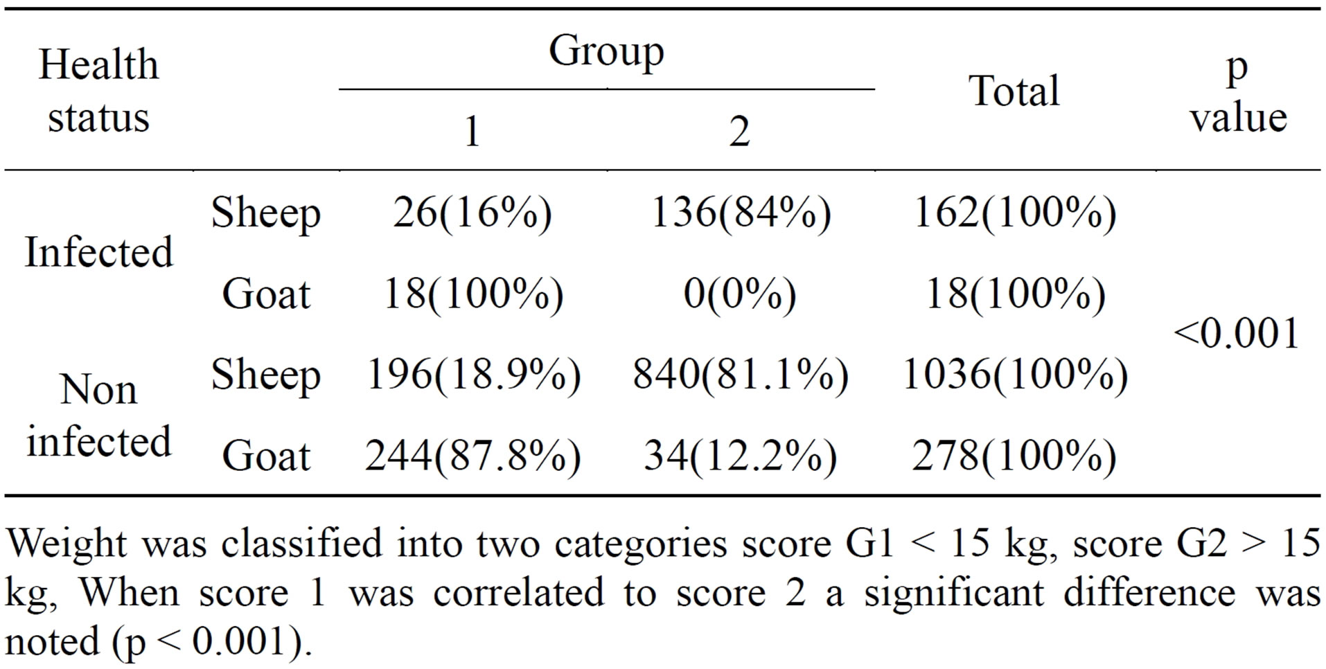
Table 4. Relationship between infection and body condition.
Weight was classified into two categories score G1 < 15 kg, score G2 > 15 kg, When score 1 was correlated to score 2 a significant difference was noted (p < 0.001).

Table 5. The relationship between age and health status in sheep and goats.
Animals were classified into two groups G1 < 12 months, G2 > 12 months.
In moderate to severe infection, the parasite may cause retarded growth and weight loss. In this study relatively young animals were slaughtered compared to other studies in the kingdom of Saudi Arabia, a significant difference was noticed among sheep, where older sheep were more exposed to the infection.
5. CONCLUSIONS
In the present study, it has been found that hydatid cysts appear mainly in liver and muscles [18] (Table 2). This is explained by the fact that the liver contains the first great capillaries sites, where Echinococcus oncosphere (hexacanth embryo) migrates and has the portal vein [10,19].
The cysts were examined to determine fertility and viability. The fertility rate was greater in sheep (66%) than goats (61%) (Table 7). The findings, 23% sterile, 66% fertile and 11% calcified cysts in sheep, may generally imply that most of the cysts in sheep are fertile which might be related to the young ages of sheep under study.
In this study it was found that more fertile cysts were harbored by sheep compared to goats, which indicates that sheep acts as the main reservoir of infection (important intermediate host). In this study most of the infected sheep were imported to be 14.9% compared to 6.3% infection among sheep of local origin which might be due to higher prevalence of hydatidosis in the origin country of the animals, whereas the infection among goats was among local goats.

Table 6. The relationship between animal source and status of infection.

Table 7. Type of hydatid cyst (sterile, fertile and calcified) in different organs of sheep and goats slaughtered in Taif abattoir.
When considering the weight of animals, it was noticed that most of the infections appear in sheep of group two with weight above 15 kg.