The Role of Bcl-2, CD10 and CD34 Expression in Differentiation between Basal Cell Carcinoma and Trichoepithelioma ()
1. Introduction
Basal cell carcinoma (BCC) is the most common cutaneous tumor, accounting for approximately 70% of all malignant diseases of the skin. It is locally aggressive and its metastasis is unusual [1] . Immunohistochemical studies support the notion that BCC originates from the basaloid epithelium of follicular bulges in the anagen hair bulbs and the follicular matrix cells [2] . Trichoepithelioma (TE) is a benign skin tumor with follicular differentiation [3] . Differential diagnosis between TE, trichoblastoma, trichofolliculoma, trichoadenoma, and BCC may be very difficult for the clinician and the pathologist [4] . Histologically, both BCC and TE are composed of nests of basaloid cells within the dermis, although these differences are distinguishable in the majority of cases [5] . CD10 is identified as the common acute lymphoblastic leukemia antigen, or CALLA [6] . CD10 may be useful for the differential diagnosis between benign tumors of cutaneous appendages originating from the hair follicle and BCC and it may solve a dilemma for the clinician and the pathologist, particularly in small and superficial biopsies [4] . Furthermore, CD10 expression can be detected in the peritumoral fibroblast-like stromal cells within the invasive area of various cancers such as prostate, breast, colorectal, and lung carcinomas [7] . CD10 expression exhibits a link with the growth rate of the cells. Its expression is increased in malignant tumors and regenerating tissues [8] . Bcl-2 is an anti-apoptotic protein residing on the outer mitochondrial membrane. It is implicated in the pathogenesis of several common cancers by inhibiting programmed cell death. In normal skin, Bcl-2 stains the majority of keratinocytes in the basal epidermis, cells of the outer root sheath (ORS), mesenchymal cells of the follicular papillae, and clear cells of eccrine glands [9] . Diffuse cytoplasmic Bcl-2 expression is reported in BCC [9] [10] , and is reported to be useful in the distinction of BCC (diffuse staining) from TE (staining of basal layer only) [11] , and in the distinction of BCC from solar keratosis (latter negative) [12] . CD34 is an intercellular adhesion protein and cell surface glycoprotein expressing in the immature hematopoietic cells and endothelial cells. CD34 is expressed in endothelial cells of the normal skin, perivascular interstitial dendritic cells of reticular dermis, around the hair follicle, and spindle cells in basal membrane zones of eccrine glands [13] -[15] .
2. Material and Methods
This study involved 32 cases; 20 cases of BCC and 12 cases of trichoepithelioma retrieved from the archives of the histopathology lab of Al Azhar University hospitals. The age, sex and site of lesions were recorded. From each paraffin block, four sections (5 micron each) were prepared for routine hematoxyllin and eosin (H&E), Bcl-2, CD10 and CD34 immunostaining. Sections stained by H&E were examined to detect pattern of growth. Sections that were immunostained for Bcl-2, CD10 and CD34 were examined microscopically to detect positively-stained tumor cells. Positive cells were considered according to (Yada et al., 2004) [16] . Reactivity of the tumor cells was analyzed for central and/or peripheral staining. Positive CD10 staining was identified as brown cytoplasmic staining with or without cell membrane staining. CD10 expression was compared with the positive control (perifollicular or perisebaceous gland area). Bcl-2 positivity was considered as diffuse cytoplasmic. Bcl-2 staining was compared with positive keratinocytes in the basal epidermis and cells of the outer root sheath (ORS). CD34 positivity was considered as cytoplasmic staining and positivity was compared with positive endothelial cells of blood vessels of the normal skin, Normal intestinal biopsy and normal tonsils were used as positive control for CD10 and CD34 respectively.
Evaluation and Statistical Analysis
All specimens were examined under a light microscope and the amount of immunupositive tumor cells and stromal cells were evaluated by using a scale of [0] to [2+] as follows: [0], negative (<10% positive cells); [1+] (10% - 50 % positive cells); [2+] (>50% positive cells).
The data were collected, tabulated, and statistically analyzed, using Statistical Package for the Social Sciences (SPSS). The Fisher exact and Chi-square tests were employed for comparison between the nominal variables, and the Mann-Whitney U test was used to compare the ordinal variables. A P value less than 0.05 were considered significant for all the tests.
3. Results
The patients with BCC were comprised of 7 females (35%) and 13 males (65%), ranging in age from 35 - 87 years. All of the BCC cases (20) were localized in the head region, 12 cases of TE 5 males (41.66%) 7 females (58.34%) ranging in age from 33 - 60 years. 11 cases were localized in the face and 1 one in nape area.
3.1. Histopathological Findings
3.1.1. TE (12 Cases)
The two major components are horn cysts of varying sizes and basaloid epithelial islands, lacking the retraction artifact typical of basal cell carcinoma. The walls of the horn cysts are formed by a few layers of cells. Some of the basophilic islands resemble follicular papillae (Figure 1).
3.1.2. BCC (20 Cases)
Nodular (8 cases; 40%) characterized by variable-sized nodules of basaloid tumor cells with elongated hyperchromatic nuclei and scant cytoplasm, peripheral palisading and clefts between tumor and surrounding stroma (Figure 2). One case was presented as pigmented nodule.
Superficial (6 cases; 30%), buds and irregular proliferations of tumor tissue attached to the undersurface of the epidermis, with clefts at the interface with the dermis.
Adenoid cystic (3 cases; 15%), show a cribriform or pseudoglandular architecture (usually with an overall nodular growth pattern) associated with mucinous stroma.
Infiltrative (3 cases; 15%), with more atypia and the infiltrating cords of basaloid cells are slender and widely infiltrative, consistent with the so-called morpheic variant of basal cell carcinoma.
3.2. Immunohistochemical Findings
Stromal and tumor cells (peripheral and/or central) expression of Bcl2, CD10 and CD34 was graded from [0] to [2+] as seen in Table 1 for BCC & Table 2 for TE.
A comparison of Bcl-2, CD10 and CD34 expression between the BCC and TE groups is displayed in Table 3 & Table 4 and that of Bcl2 in both lesions in Table 5, and that of CD10 in both lesions in Table 6, and that of CD34 in both lesions in Table7
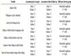
Table 1. Expression of CD10, Bcl-2 and CD34 in BCC.
Table 2. Expression of CD10, Bcl-2 and CD34 in trichoepithelioma.
Table 3. Expression of CD10, Bcl-2 and CD34 in BCC.
*P-value < 0.05 is significant. So, Bcl2 staining is significant in tumor cells and insignificant in stromal cells, CD34 staining is significant in tumor cells and insignificant in stromal cells, and CD10 staining is insignificant in both tumor and stromal cells.
Table 4. Expression of CD10, Bcl-2 and CD34 in trichoepithelioma.
So, Bcl2 staining is significant in tumor cells and insignificant in stromal cells, CD34 staining is significant in tumor cells and stromal cells, and CD10 staining is insignificant in tumor cells and stromal cells.
Table 5. Expression of Bcl-2 in BCC vs trichoepithelioma.
*P-value < 0.05 is significant, so, Bcl2 staining is significant in tumor cells of both BCC and TE, and insignificant in stromal cells of both BCC and TE.
Table 6. Expression of CD10 in BCC vs trichoepithelioma.
CD10 staining is insignificant in both BCC and TE.
We noted that Bcl-2 immunoreactivity in tumor cells was strong in both BCC and TE, but it is stronger in basal layer in TE (Figure 3) and diffuse in BCC (Figure 4 & Figure 5) and. CD10 immunoreactivity was strong in peritumoral stromal cells and negative in tumor cells of TE (Figure 6) and stronger in basaloid cells of BCC (Figure 7), and CD34 immunoreactivity was stronger in stromal cells of TE (Figure 8) than BCC (Figure 9).
Table 7. Expression of CD34 in BCC vs trichoepithelioma.
CD34 staining is significant in tumor and stromal cells of TE, and significant in tumor cells of BCC.

Figure 1. TE: Aggregates of basaloid cells in dermis with peripheral palisading, no clefts (H&E ×150).

Figure 2. BCC: infiltrating nodules with surrounding cleft artifacts (H&E ×135).

Figure 3. TE: stronger Bcl-2 staining of basaloid tumor cells, (DAB ×235).

Figure 4. BCC: showing diffuse Bcl-2 staining of tumor cells (DAB ×235).

Figure 5. BCC: showing diffuse Bcl-2 staining of tumor cells (DAB ×360).

Figure 6. TE: CD10 positive stromal cells and negative tumor cells (DAB ×135).

Figure 7. BCC: CD10 stronger staining of peripheral cells of nodules (DAB ×235).

Figure 8. TE: strong CD34 staining of stromal cells, and negative tumor cells (DAB ×360).

Figure 9. BCC: CD34 positive stromal cells and BV, and negative tumor cells (DAB ×360).
4. Discussion
In the present study, we examined 20 BCC and 12 TE for Bcl-2, CD10 and CD34 expression to see the immunostaining pattern of BCC and TE and if these makers might aid in the differentiation between the two tumors.
The Bcl-2 positivity was seen in 60% and 100% of BCC stromal cells and tumor cells respectively, While there was 33.3% and 100% Bcl-2 expression in TE stromal cells and tumor cells respectively, and the pattern of staining is diffuse in BCC and stronger in basal layer in TE.
These findings are in agreement with some studies which stated that Bcl-2 diffusely stains the tumor nests in BCC while it stains the outermost cell layers in trichoepithelioma (Verhaegh et al., 1997 [17] , and Smoller et al., 1994).
CD10 positivity was seen in 40% and 55% of BCC stromal cells and tumor cells respectively while there was 58.3% and 41.6% CD10 expression in TE stromal cells and tumor cells respectively.
CD10 immunoreactivity in BCC, basaloid cells are strongly positive while in TE CD10 was strongly positive in peritumoral stromal cells with patchy staining of basaloid cells.
These findings are nearly in agreement with Sengul et al., (2010) which showed condensation of CD10-positive stromal cells around basaloid nests, which was statistically significant in differentiating TE from BCC. Conversely, CD10-positive basaloid cells were seen predominantly in BCC. No BCC cases demonstrated stromal expression alone in that study. The expression of CD10 by peritumoral stroma alone favored a diagnosis of TE, whereas staining of basaloid cells supported a diagnosis of BCC.
Wagoner et al., 2007 showed strong CD10 expression in the tumor cells of superficial BCC.
CD34 positivity was seen in 70% of BCC stromal cells, and 83.3% of TE stromal cells, but tumor cells in both tumors showed no immunoreactivity.
The findings in this study, are in agreement with Sengul et al., (2010) which showed stromal expression of CD34 for 25 (83.3%) of 30 cases in benign tumors of cutaneous appendages originating from hair follicle (BTCOHF) and 9 (30%) of 21 (70%) in BCC. While stromal expression of CD34 for BTCOHF was observing just the adjacent to the tumor islands; it was not like that for BCC. There was CD34 expressions in the surrounding stromas of BCCs with the adjacent zones which were not stained with CD34.
Illueca et al., 1998 [18] showed the usefulness of CD34 by showing the lack of CD34 expression by tumor stroma in BCC, but positive in TE.
The findings of this study are different from findings of Kirchmann et al., (1994) [19] which stained 19 cases of BCC and 16 cases of TE with CD34 in their study. They observed that while the spindle-shaped cells surrounding the islands of trichoepithelioma cells were focally strongly positive for CD34; surrounding the nests of tumor cells were negative in all basal cell carcinomas. So they suggested that, CD34 staining pattern differentiates between trichoepithelioma and basal cell carcinoma.
The findings of this study are different from findings of Naeyaert et al., (2001) which compared the CD34 staining patterns of TE with (fibroepithelioma) and nodular BCC. They also did not detect the peritumoral stromal expression of CD34 in those variants of BCC.
5. Conclusion
In conclusion we can suggest that bcl-2, CD10, and CD34 are useful markers in differentiation between BCC and TE. Positive immunostaining for bcl-2 tends to be diffuse in BCC, whereas it is peripheral in trichoepithelioma. Positive immunostaining for CD10 in stromal cells around basaloid nests favors TE over BCC. In addition, CD34 is found to be more positive in the stroma of trichoepithelioma than BCC and absent in tumor cells of both tumors. These findings may prove to be of diagnostic help in distinguishing borderline cases, and also offer some possible explanations for the biological differences between these neoplasms.