Clinical Profile and In-Hospital Outcome of Patients with Right Ventricular Myocardial Infarction ()

1. Introduction
Right ventricular myocardial infarction (RVMI) as assessed by various diagnostic methods accompanies inferior-posterior wall myocardial infarction in 30% to 50% of patients. Recognition of the syndrome of right ventricular involvement is important as it defines a significant clinical entity, which is associated with considerable immediate morbidity and mortality and has a well-delineated set of priorities for its management.
Looking into the pathogenesis of the disease, right and left ventricles differ markedly in their anatomy, mechanics, loading conditions, and metabolism, and therefore it should not be surprising that they have strikingly different oxygen supply and demand characteristics and thus manifest disparate responses to ischemic insults [1] -[3] . Patient may be clinically present with hypotension, elevated jugular venous pulse (JVP), and occasionally shock, all in the presence of clear lung fields [4] . The ST-segment elevation of ≥0.1 mV in the right precordial leads (V4R) of the Electrocardiograph (ECG) is a readily available electrocardiographic sign used for diagnosis of RVMI. Hence ECG has always been the first choice of investigation to detect RVMI. The method is easy and cost-effective too.
Patients with inferior wall myocardial infarction (IWMI) who have right ventricular myocardial involvement appear to have a worse prognosis than those who do not have right ventricular involvement [5] -[7] . Since there is scarcity of literature regarding epidemiology of clinical profile as well as in-hospital outcomes of patients with RVMI in the Indian population, this study is carried out with a goal of identifying RVMI in our hospital setting.
2. Methods
2.1. Study Methodology
A prospective observational study was conducted at the department of cardiology, medical college, Kottayam during December 2007 to November 2008. The protocol of the study was approved by institutional ethics committee of the hospital before the commencement of the study. Informed consents were obtained from all the patients enrolled in the study.
All the patients with definitive evidence of acute IWMI as proved by 12-lead ECG with right precordial leads and window period of chest pain less than 24 hours were considered in our study. It included a total of 200 patients, 100 consecutive patients with only IWMI and 100 patients of right ventricular involvement in IWMI. Patients with anterior wall myocardial infarction were excluded from the study.
A detailed case history was taken and careful physical examination was done with special reference to JVP, hypotension, S3/S4 and cardiac murmur. ECG monitoring was done during the stay in the intensive cardiac care unit for the identification of arrhythmia and conduction blocks. Routine serum investigations like random blood sugar, serum lipid profile, renal function test, blood routine, cholesterol along with cardiac enzymes and serum glutamic oxaloacetic transaminase were done at the time of admission. ECG was monitored in all the cases.
Acute myocardial infarction was treated with or without thrombolytic therapy. During the study period primary percutaneous coronary intervention facility was not available in our setup. In cases with RVMI who were haemodynamically compromised, volume loading was done using normal saline (and inotropes in unresponsive cases). Arrhythmia was treated accordingly.
2.2. Statistical Analysis
Descriptive statistical analysis was performed to express the results of the study. We applied Student T test for continues variable, and for categorical Chi Square and Fisher Exact test.
3. Results
The results obtained from the study are shown in Table 1.
Analysing the gender of the patients, males mostly predominated both the classes of patients. Average age of the patient was found to be 52 years in patients with IWMI and 64 years in the other class.
Smoking was found to be the most significant risk factor. Ninety percentages of patients with IWMI, and 96% of RVMI were found to be smokers.
Thirty-eight out of 100 patients of RVMI died because of hypotensive shock whereas cardiogenic shock was
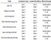
Table 1. Clinical and echocardiographic changes in patients with IWMI and RVMI.
responsible for the death of 20 patients suffering from RVMI. In patients with IWMI, hypotension was responsible for the death of 8 out of 100. Two out of 100 patients suffering from IWMI died from cardiogenic shock. Hence we conclude hypotension as a sign of clinical presentation in patients with RVMI is more jeopardizing. Among electrical disturbances primary ventricular tachycardia was frequently seen in patient with RVMI. The mortality rate was found to be 12% in patients with IWMI and 28% in patients with RVMI. From the above study, it was found that mortality rates are quite high in the patients with RVMI.
4. Discussion
Based on early experiments of right ventricular performance, it was felt for many years that right ventricular contraction was unimportant in the circulation and that, despite loss of right ventricular contraction, pulmonary flow could be generated by a passive gradient from a distended venous system and active right atrial contraction. However, recognition of the profound hemodynamic effects of right ventricular systolic dysfunction became evident during the 1970s with the description of severe RVMI, resulting in severe right heart failure, clear lungs, and low-output hypotension despite intact global left ventricular systolic function [8] .
It has been very well noted in the study that significant RVMI nearly always occurs in association with acute transmural inferior-posterior left ventricular myocardial infarction, and the right coronary artery (RCA) is always the culprit vessel [9] [10] , typically a proximal occlusion compromising flow to one or more of the major right ventricular branches.
Acute RVMI is associated with higher in-hospital morbidity and mortality related to life-threatening hemodynamic compromise and arrhythmias during acute occlusion and abruptly with reperfusion, complications which have implications for interventional management [11] .
In 1981, the sensitivity and specificity of ST-segment elevation in the right precordial lead V4R as an early indicator of right ventricular infarction were examined in a consecutive series of 110 patients admitted for acute inferior myocardial infarction. Some other studies have also showed this type of observations [12] [13] . The sensitivity was 82.7%, the specificity was 76.9% and the positive predictive value was found to be 70% in 58 patients with RVMI. Because of its simplicity and its high sensitivity and specificity, recording of V4R should be an intrinsic part of the early evaluation and electrocardiographic examination of acute IWMI [14] . In combination to the ECG, the triad of hypotension, elevated jugular venous pressure and clear lung fields have been recognised as markers of RVMI in IWMI [8] [15] [16] .
Thirty-two patients with RVMI were reported to have hypotension in our study. On the other hand, only 8 patients of IWMI were found to have a hypotensive condition.
Elevated JVP is usually present with a Y descent greater than or equal to X descent, implying that the right ventricle is poorly complaint. Using a haemodynamic gold standard, Italia et al. found elevated JVP to be 88% sensitive, yet only 69% specific for RVMI in IWMI. Caution must be exercised in relying on such findings since they are readily masked by volume depletion and emerge only after volume loading [17] . 52% of the study population suffering from RVMI in our study showed this sign.
Kussmaul’s venous sign (distension of the jugular vein on inspiration), has also been shown to be highly sensitive and specific for RVMI. Cintron and associates detected Kussmaul’s sign in 16 of 45 patients with inferior or posterior transmural infarctions (not seen with anterior or lateral events) [18] . Out of 16, 9 had heamodynamically significant right ventricular involvement in the study done by Cintron et al. [18] . In one series, 11 of 46 patients with Kussmaul’s sign had a right sided 4th heart sound and 4 had right sided 3rd heart sound [19] . In our study, 42 patients with RVMI showed this sign, whereas no patient of IWMI showed a positive Kussmaul’s sign.
Patients with acute RVMI are at an increased risk for high-grade atrio-ventricular (AV) block, bradycardia and hypotension without AV block. Recent findings now document that during acute coronary occlusion, bradycardia hypotension and AV block are far more common in patients with proximal RCA lesions inducing right ventricle and left ventricle inferior-posterior ischemia [10] .
In the present study, average age of incidence was 64 years in RVMI group and 52 years in the group which consisted of patient with RVMI along with IWMI. S. Khan et al. reported 100 cases of IWMI in which 86% were males and 14% females [20] . In our study, males predominated in RVMI and non-RVMI groups, 62% and 60% respectively. As reported in the study by S. Khan, in-hospital mortality (23.5%) was higher in RVMI group than isolated IWMI (18.1%) [20] . Other major complications were also higher in RVMI group than isolated IWMI. Similar observation was obtained in our study which reported mortality rate of 28% in patients with RVMI and 12% in patient with IWMI. Right ventricular infarction was found in approximately one-third of IWMI. RVMI was associated with considerable morbidity and mortality, and its presence defines a higher risk subgroup of patients with IWMI.