1. Introduction
Synovial Sarcomas (SS) are a group of Soft Tissue Sarcomas (STS) affecting mainly young adults. The most common site of occurrence is in extremities [1].
Primary Synovial Sarcoma (PSS) of the kidney is a recently described entity [2].
Fewer than 50 cases of primary renal synovial sarcoma are reported to date [3].
This tumor presents a diagnostic dilemma because it is quite difficult to differentiate it from other renal neoplasms, such as metastatic sarcoma, renal cell carcinoma with sarcomatoid differentiation, which may have similar histological features.
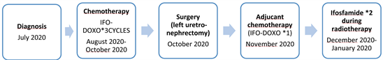
In the present report, we present the case of a 13-year-old girl who was diagnosed with primary renal synovial sarcoma.
2. Case Report
A 13-year-old female was presented with a mild left flank pain of one-week duration, with no associated history of hematuria or any other systemic symptoms.
Renal ultrasound showed a large left upper polar renal mass 16 × 13 × 9 cm, hypoechoic, homogeneous, well limits, presenting rich vascularization on color Doppler.
The diagnosis of nephroblastoma has been suggested.
Computed tomograpgy of the abdomen and pelvis showed a left retroperitoneal mass probably renal origin measuring 14 × 10 × 11.5 cm associated with retroperitoneal adenomegaly.
A CT scan of the chest did not reveal metastatic disease (Figure 1).
The patient underwent a kidnet biopsy which revealed a histological and immunohistochemical monophasic spindle cell synovialosarcoma of left kidney.
On immunohistochemistry, the tumor cells showed an intense and diffuse nuclear activity for TLE1, focal positivity for cytokeratin AE1/AE3.
WT1 was focally positive but this positivity is not specific (Figure 2).
According to these findings, a diagnosis of primary monophasic SS of the kidney was made.
A complete tumor resection with node dissection cannot be obtained, the patient was referred to our department of pediatric oncology in sousse for the neoadjuvant chemotherapy.
She received chemotherapy with Doxorubicin 37.5 mg/m2/day for 2 days and Ifosfamide 3 g/m2/day for 3 days every 21 days for a total of 3 cycles according to EpSSG NRSTS 2005 protocol with good tolerance.
2 weeks after the chemotherapy, the patient had a CT of the abdomen and pelvis which showed a decrease in size of the heterogeneous left renal mass of 8 cm (vs. 14 cm) (estimated at 42%), clear regression of the effect of mass on the anterior structures previously described with, however, persistence of intimate contact with the tail of the pancreas and the splenic vein.
No retro-peritoneal adenomegaly of significant size.
No new lesion (Figure 3).
The patient was transferred to pediatric surgery department, she underwent exploratory laparotomy that involved the resection of a 10 cm mass, with a left uretro-nephrectomy (Figure 4).
![]()
Figure 1. Coronal section of a computed-tomographic scan of the abdomen and pelvis, showing a left retroperitoneal mass with fine calcifications and few hypodense areas of necrosis.
![]()
Figure 2. Tumor composed of spindle cells arranged in intersecting fascicles alternating with hypocellular areas.
![]()
Figure 3. CT image showing marked decrease in tumor size.
![]()
Figure 4. Macroscopically, the tumor mesarures 10 cm and shows fibro-necrotic changes secondary to chemotherapy.
Histology revealed post-chemotherapy residue of monophasic spindle cell synovialosarcoma of the left kidney.
The tumor measures 10 cm long and shows fibro-necrotic changes secondary to chemotherapy estimated at 80% of the tumor volume.
Absence vascular embolus of the hilum vessels.
After recovering from surgery, the patient received one cycle of chemotherapy (Doxorubicin 37.5 mg/m2/day for 2 days and Ifosfamide 3 g/m2/day for 3 days) and 2 cycles of chemotherapy (Ifosfamide alone at 3 g/m2/day for 2 days) concomitantly to radiotherapy. She received 45 Gy (1.8 Gy/d) at the 9th week, concomitantly to 4th and 5th cycles with a good tolerance.
After 6 months the patient came for follow up and she was doing well.
3. Discussion
Monophasic synovial sarcoma of the kidney is an extremely rare tumor and less than 50 cases have been described in the literature.
It was first reported in 1999 by Faria et al. [4].
Synovial sarcoma usually involves adolescents and young adults; however, the age at presentation ranges from 17 to 61 years. Diagnosis is difficult due to the rarity of the tumor and its similar presentations as compared to other renal tumors. Differential diagnosis includes Adult Wilms tumor, transitional cell carcinoma, renal cell carcinoma and hemangiopericytoma, congenital mesoblastic nephroma, and primitive neuroectodermal tumor [5].
The patients commonly present with flank pain and/or hematuria. No clinical feature or imaging modality is diagnostic. The CT scan usually reveals a heterogeneously enhancing renal mass and the confirmation of diagnosis is by molecular and cytogenetic analysis. Rarely is the presentation at an advanced stage with caval thrombus and/or metastasis. To the best of our knowledge, approximately three cases of SS with the caval thrombus have been reported previously. In 2007, Tornkvist et al. [6] reported six cases of metastatic disease.
Primary synovial sarcoma occurs in two forms: Biphasic and Monophasic. The primary biphasic synovial sarcoma contains both glandular elements and spindle epithelial cells. The primary monophasic synovial sarcoma is composed of only spindle cells [7].
There are no established guidelines regarding management of this tumor given the limited number of cases reported. Primary surgical treatment is considered to be the treatment of choice; prognosis is poor with this treatment alone. Surgical resection is the mainstay, although surgery alone has a poor prognosis. Reports of sensitivity of this tumor to ifosfamide- and doxorubicin-based chemotherapy do exist but no clear guidelines are available regarding adequate treatment of this rare entity [8].
Radiotherapy is also effective in presence of local spread as it was in our patient [9].
4. Conclusions
In conclusion, primary SS of the kidney is an extremely rare disease and preoperative diagnosis is difficult in the absence of specific clinical or imaging findings.
Its diagnosis is based on morphological and molecular studies.
The combination of surgery and chemotherapy has shown positive results. Particularly, the use of Ifosfamide and Doxorubicin is as standard chemotherapy to induce complete remission.
Radiotherapy can be useful as adjuvant therapy in presence of local spread.