Correlation of Hamstring Tendon Size in Pre-Operative MRI Measurement with Intra-Operative Graft Size in Primary Anterior Cruciate Ligament (ACL) Reconstruction ()
1. Introduction
The use of hamstring autograft for anterior cruciate ligament (ACL) reconstruction was first reported in 1934 with the use of the semitendinosus tendon. Advancements in surgical techniques have led surgeons to use 2 hamstring tendons for ACL autograft reconstruction with both the semitendinosus (ST) and gracilis tendons (GT) in a 4-stranded construct. A retrospective review of patients who underwent ACL reconstruction using quadrupled semitendinous and gracilis autografts to determine the relationship between graft failure rate and predictor variables have found that Hamstring graft diameter of 8.0 mm or more was associated with significant reduction of risk in graft failure rate [1] [2] .
Concern regarding the potential of a higher failure rate with use of thinner hamstring grafts has led some investigators to the practice of adding allograft tissue to increase the overall graft diameter when thickness of harvested tendons is deemed insufficient [3] . Others prefer to increase the number of times the harvested hamstring tendons are folded to obtain a thicker, albeit shorter, final graft. But the ability to fold a tendon on itself more than once to increase graft thickness requires the harvested tendon to be long enough to allow that. Indeed, a recent description of the use of a 5-stranded graft for ACL reconstruction by tripling the ST indicated that harvested tendon needed to be at least 21 cm long to yield a graft of sufficient length [4] .
Both imaging and anthropometric patient-specific methods have been shown to be accurate and consistent in predicting hamstring graft diameter. Anthropometric data are similarly useful for hamstring graft size prediction. Height was the most common predictor of larger grafts [5] [6] . Overall, the quality of the correlations with graft diameter obtained by any parameter in previous studies was weaker than that of the correlations obtained by MRI cross-sectional area (CSA). Because most surgeons currently obtain an MRI study of the knee before ACL-R and many software packages contain tools to measure CSA, this method of prediction may be added to routine preoperative planning with a small addition in the protocol [7] [8] .
There are multiple studies done using imaging (MRI and CT scan) to predict and correlate hamstring graft size. In addition, all used similar methods of predicting graft diameter based on the combined CSA of the semitendinosus and gracilis tendons. This was performed at similar locations along the knee joint and was obtained by software analysis with a region-of-interest tool based on the initial protocol developed by Bickel et al. [9] Other than the earliest attempt to predict graft diameter by Yasumoto et al. in 1997, the only study based on CT, all 5 subsequent studies found significant Pearson correlation coefficients between CSA and intraoperative graft diameter [10] .
The aim of this study is to assess the correlation of ACL graft size with the preoperative MRI measurement of both the CSA and tendon-only length of the Hamstring tendon (semitendinosus and gracilis). Thus, it may help surgeons to anticipate the needs for graft augmentation should the final graft size be smaller than expected.
2. Material and Methodology
This is a retrospective study of patients who underwent ACL reconstruction at Hospital PakarSultanah Fatimah (HPSF), Muar, from January 2019 to December 2022. We have identified all cases of 18 year old and above, with primary ACL reconstructions using hamstring autograft, for which preoperative MRI was done. The final Hamstring graft size (triple loop graft) from operative notes was extracted. Patients with previous knee surgery other than diagnostic arthroscopy, multi-ligament injury, or fracture around the ipsilateral knee, and patients with amputated graft during the surgery were excluded.
Patients demographic data and the date of surgery were recorded in the data collection sheet. Final Hamstring graft size (6-strands; diameter and length) were identified from the operative notes.
All the MRI from selected patients were read by a single Radiologist and recorded in the Data Collection Sheet. The CSA of both Semitendinosus and Gracilis tendon were taken at the widest point of the distal femur close to the joint line, as this is the point where both tendons are most rounded and oval in shape. Because the MTJs and tibial insertions may not be visible on the same image slice, for each tendon the distal-most aspect of the MTJ (confirmed in at least 2 planes) were digitally marked and this point translated to a fat-suppressed, PD FSE sequence sagittal image depicting the tibial insertion, which were then also be digitally marked. The straight-line distance between these 2 points will be reported as the tendon-only length of each hamstring.
The data were summarised with descriptive statistics based upon the distribution of the data. Correlation between MRI measurements and the actual graft sizes were tested using Pearson’s correlation test. The prediction of actual graft size (the outcome/dependent variable) using the MRI CSA measurements as the predictor/independent variable will be modelled using simple linear regression.
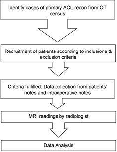
a) Flow Chart of the Study Methodology
3. Results
Of 105 patients with primary ACL reconstruction done between January 2019 to December 2022, only 41 patients were included in this study. 26 (63.4%) were male and another 15 (36.6%) were female and mean age of the patients was 28 year old. 97.6% were Malay patients. Majority of the cases had sustained sports-related injury (73.2%) and 17.1% had involved in motor-vehicle accidents, and another 9.7% were domestic injuries and othersas shown in Table 1.
In general, the correlation between MRI-measured Hamstring thickness and intra-operative graft diameter were good, but not excellent (p = 0.048) (Table 2). We found that the minimum combined diameter of ST and gracilis tendon of 11.5 mm will produce graft of 7.0 mm, and maximum reading of 26.7 mm in this study will produce graft as big as 11.0 mm. Whilst the mean pre-operative combined diameter of 17.0 will results in graft diameter of 8.3 mm. The mean length of ST and gracilis tendon of 11.0 cm and 8.9 cm in pre-operative MRI will give a triple-loop graft of 9.0 cm (Table 3). In Figure 1, the graft shows the agreement between the pre-operative Hamstring diameter and and the intra-operative final graft (triple-loop) diameter.
In this study, a Pearson correlation coefficient was computed to assess the relationship between height and intra-operative diameter and there was positive correlation between the two variables (p < 0.001) (Table 4). It is also shown in Figure 2, that the taller the patients, there’s also high possibility to get bigger graft.
4. Discussion
The hamstring tendon graft diameter plays an important role in the outcome of ACL reconstruction [11] [12] . Unlike patellar tendon or quadriceps tendon autografts, in which a consistent diameter can be obtained, the diameter of a hamstring
![]()
Table 1. Demographic profile of patients.
![]()
Table 2. Agreement of diameter between pre- and intra operative Item Statistics.
Average measuring of diameter from two different time of operation is poor reliability based on recommendation given by Koo and Li (2016). The intraclass correlation (ICC) by using two-way mixed model with type consistency, the reliability based on average measure was 0.41 (95% CI: −0.10, 0.69).
![]()
Table 3. Clinical outcome pre- and intra-operative.
![]()
Figure 1. Bland Altman graph showing agreement between pre op diameter and intra op diameter.
![]()
Table 4. Correlation between height and intra-op diameter.
A Pearson correlation coefficient was computed to assess the relationship between height and intra op diameter. There was a positive correlation between the two variables, r = 0.685, n = 41, P =< 0.001, which suggests moderate correlation (Munro, 2000).
![]()
Figure 2. Relationship between height and Intra op diameter.
tendon autograft is highly variable and sometimes unpredictable. In this study, we found a good correlation between pre-operative MRI Hamstring tendon thickness (CSA) with the triple-loop (six-strand) graft for ACL reconstruction, hence MRI is a good tool to predict the graft size for the surgery.
This study has limitation by the fact that all measurements were made by a single radiologist. Therefore, interobserver variability was not evaluated. Interobserver variability will be evaluated as a prospective arm of this measurement protocol. Nevertheless, the radiologist was shown to have excellent intra-observer reliability throughout the study, indicating consistency in the measurements. As with any retrospective study, there is concern of possible selection bias. However, each patient’s record was reviewed at the time of data collection to ensure that only patients who met our criteria were enrolled. Finally, we did not attempt to correlate our findings on graft size with knee function, return to sport, or objective measures such as range of motion (ROM) or stability, as these outcomes were beyond the scope of our study.
Besides the CSA of Hamstring tendons, we also have assessed the length of each tendon in the MRI. The correlation was made to the final graft length, which was also triple-loop of the tendon. The limitations of this study include the lack of documentation of the total length of each tendon intra-operatively. Furthermore, attempting to measure the entire length of the ST or gracilis on routine MRI scans of the knees is frustrated by the fact that the imaged region often does not cover the entire length of these tendons.
A study done by Salman et al. [13] showed that height, weight, thigh length and circumference all demonstrated a moderately positive correlation with graft size within this review. Such anthropomorphic measurements can be considered surrogate markers for muscular development, both in relation to cross-sectional area and axial muscular length and thus can be considered more relevant markers to base potential graft size upon. This results also supported by another study done by Moghamis [14] where he found positive correlation with the hamstring graft length and diameter in male patients. It can also be seen in our study where there is positive correlation with the patients height and the final Hamstring graft. However, the anthropometric measurement alone is sometimes can mislead the surgeon in predicting the graft size.
5. Conclusion
These results showed good correlation with the pre-operative MRI measurement of Hamstring tendon and the intra-operative graft size in ACL reconstruction. There was also positive correlation with the patients’ height and the final graft size. Hence, use of MRI and patients anthropometric may be used to predict the Hamstring graft size in ACL reconstruction.