 Journal of Cancer Therapy, 2013, 4, 466-474 http://dx.doi.org/10.4236/jct.2013.43A057 Published Online March 2013 (http://www.scirp.org/journal/jct) MicroRNAs as Targets and Tools in B-Cell Lymphoma Therapy Kalman Szenthe1, Katalin Nagy2, Krisztina Buzas3,4, Hans Helmut Niller5, Janos Minarovits3* 1RT-Europe Nonprofit Research Center, Mosonmagyarovar, Hungary; 2Department of Oral Surgery, Faculty of Dentistry, University of Szeged, Szeged, Hungary; 3Department of Oral Biology and Experimental Dental Research, Faculty of Dentistry, University of Szeged, Szeged, Hungary; 4Institute of Biochemistry, Biological Research Center of the Hungarian Academy of Sciences, Szeged, Hungary; 5Department of Medical Microbiology and Hygiene, University of Regensburg, Regensburg, Germany. Email: *minimicrobi@hotmail.com Received January 23rd, 2013; revised February 25th, 2013; accepted March 5th, 2013 ABSTRACT MicroRNAs (miRNAs) are posttranscriptional regulators fine-tuning the level of most messenger RNAs (mRNAs) and proteins in mammalian cells. Their expression is dysregulated in neoplastic cells and upregulated or downregulated miRNAs play an important role in tumorigenesis. Changes in the miRNA transcriptome appear to be suitable markers for the differential diagnosis of various B-cell lymphoma types and there are therapeutic attempts to normalize the ex- pression level of key cellular miRNAs involved in lymphomagenesis. In this review we wish to outline the most recent developments in the application of sophisticated, chemically modified antisense oligonucleotides and their nanoparticle complexes to suppress oncogenic miRNAs. These advances form the basis of a new therapeutic approach that may complement current protocols for B-cell lymphoma therapy. Anti-cytokine therapy aiming at the removal of cytokines that activate key oncomirs, and switching on silenced tumor suppressor miRNAs by epigenetic drugs might also be considered, on the long run, in the treatment of well defined B-cell lymphoma types. Keywords: MicroRNAs; B-Cell Lymphoma Therapy; Antisense Oligonucleotides; Anti-Cytokine Therapy; Epigenetic Drugs 1. Introduction Recently, microRNAs (miRNAs) emerged as a new class of regulators of a wide variety of physiological processes [1]. miRNAs usually downregulate messenger RNA (mRNA) levels and protein levels by binding to comple- mentary regions on their target mRNAs. miRNA-mRNA association may either facilitate mRNA breakdown or block mRNA translation. Dysregulated expression of certain miRNAs plays a role in the development of malignant neoplasms [2-4]. Increased level of several miRNAs, called oncomirs, fa- cilitates oncogenesis and tumor progression. Other mi- RNAs act similarly to the products of tumor suppressor genes: their reduced expression may result in uncontrol- led cell proliferation and altered cellular behaviour. 2. miRNAs as Pleiotropic Posttranscriptional Regulators in Normal and Neoplastic Cells 2.1. Biogenesis of MicroRNAs miRNAs are transcribed in most cases by RNA poly- merase II (Pol II) from independent transcription units or they are processed from introns of transcripts (for review see [1,3]). Typically the primary Pol II product (pri- miRNA) is processed in the nucleus by the RNase III Drosha resulting in a stem and loop structure called pre- miRNA. The approximatively 70 nucleotide long pre- miRNA molecule is carried to the cytoplasm by Exportin 5, a nuclear export protein. As a next processing step, DICER1, a cytoplasmic RNAse III enzyme removes the loop of the pre-miRNA and the resulting double stranded RNA is loaded onto an Argonaute (Ago) protein, a com- ponent of the RNA-induced silencing complex (RISC). One strand of the approximately 22 nucleotide long du- plex RNA represents the guide strand or mature miRNA that remains associated with Ago, whereas the other strand is degraded. As few as 6 nucleotides of the mature single stranded miRNA (nucleotides 2 - 7) form the “seed region” that plays a major role in selecting the 3’ untranslated region (3’ UTR) of the target mRNA. Be- cause the seed region, where full complementarity is re- quired with the target mRNA for RISC function, is short, a miRNA may potentially recognize multiple mRNA tar- gets. It is also worthy to note, however, that the very *Corresponding author. Copyright © 2013 SciRes. JCT  MicroRNAs as Targets and Tools in B-Cell Lymphoma Therapy 467 same 3’ UTR may bind more than one miRNA species (reviewed in [1,3]). 2.2. Dysregulation of miRNA Expression in B-Cell Lymphomas The pattern of miRNA expression in neoplastic cells dif- fers from the miRNA transcriptome (miRNAome) of their normal counterparts (for review see [3]). In addition, the expression signatures of miRNAs appear to be suit- able markers for the differential diagnosis of various ma- lignant tumors including the different B-cell lymphoma types [5]. Some of the differentially expressed miRNAs apparently play an important role in the initiation and maintenance of the malignant behaviour, which implies that overexpression or inhibition of key microRNAs may revert the malignant phenotype. The best characterized miRNAs most frequently upregulated or downregulated in the major B-cell lymphoma types are summarized in Tables 1 and 2 (for a comprehensive review see also [4]). Tables 1 and 2 also show their target mRNAs and puta- tive role in lymphomagenesis. It is important to note that—depending on the cellular context—the very same miRNA may either facilitate or inhibit tumorigenesis. Thus, miR-155, a multifunctional regulator molecule up- regulated in diffuse large B-cell lymphomas (DLBLs) and Hodgkin lymphomas acted as an oncomir in a trans- genic mouse model, where its ectopic expression resulted in pre-B cell proliferation and lymphoblastic leukemia/ lymphoma [6-8]. All these changes were attributed to a release of the interleukin-6 (IL-6) pathway from its in- hibitory control by miR-155 [9]. In gastric cancer cells, however, miR-155 was downregulated and its overex- pression suppressed cell migration and invasion. Thus, in gastric cancer cells miR-155 acted as a tumor suppressor targeting SMAD2, a component of the transforming growth factor beta (TGFβ) signalling pathway [10]. In addition, Babar et al. observed that although induction of high levels of miR-155 in a tetracycline-controlled knock- in mouse model caused the development of disseminated pre-B cell lymphoma, highly elevated miR-155 expres- sion in the brains of the same mice was insufficient to induce brain tumors [11]. 2.3. Attempts to Normalize the Expression of Key Cellular miRNAs in Various B-Cell Lymphoma Types As illustrated above, miR-155 has emerged as a pleio- tropic regulator of numerous biological processes. Thus, it became a logical therapeutic goal to attenuate its on- cogenic functions to complement, on the long run, cur- rent protocols for lymphoma treatment. High affinity binding of anti-miR oligonucleotides to distinct micro- RNAs may sequester their target without causing its de- gradation, whereas a lower-affinity binding promotes miRNA degradation [12]. Early in vivo attempts of se- quence specific silencing by short hairpin RNAs failed due to the activation of the immune system, toxic off- target effects, and saturation of the endogenous micro- RNA pathway (reviewed in [13,14]). However, more so- phisticated, chemically modified antisense oligonucleo- tides including locked nucleic acid (LNA) oligonucleo- tides, peptide nucleic acids (PNAs) conjugated either to a cell-penetrating peptide or polylysine, and cholesterol- conjugated RNAs called “antagomirs” proved to be ef- fective inhibitors of microRNAs both in vitro and in vivo [14-18]. Fabani et al. reported that a peptide nucleic acid (PNA) oligonucleotide containing four D-lysine residues to ensure proteolytic stability and enhanced cellular uptake could inhibit miR-155 both in vitro and in the spleens of experimental mice receiving 50 mg PNA/kg/ day for 2 days [16]. To explore the advantages of the nanoparticle delivery systems that permit targeting of miRNA inhibitors to se- lected cells and tissues, anti-miR-nanoparticle complexes were constructed and tried successfully in clinically ac- ceptable and therapeutically affordable doses in mice [11, 19,20]. Babar et al. demonstrated that in spite of the po- tential difficulties of cellular internalization, antisense pe- ptide nucleic acids encapsidated into biodegradable poly (lactic-co-glycolic acid) (PLGA) particles coated with penetratin inhibited miR-155 and decreased the growth rate of pre-B-cell tumors in a knock-in mouse model [11]. Although penetratin facilitates uptake into various cell types, it was observed that the systematically adminis- tered nanoparticles showed preferential targeting to tu- mor tissue. miR-155 is indispensable for the development of an optimal T cell dependent antibody response and controls T helper cell differentiation by regulating cytokine pro- duction [21]. miR-155 expression leads to elevated tumor necrosis factor alpha (TNFα) levels whereas TNFα in- duces miR-155 expression. In a subgroup of diffuse large B cell lymphoma, called activated B cell-like or non- germinal centre (ABC or non-GC) DLBC, augmented miR-155 expression was found due to an increased TNFα production. The high miR-155 level could be reduced in non-GC DLBC cell lines cultured in vitro by adding eternacept, a soluble TNFα receptor or infliximab, a neu- tralized humanized monoclonal antibody against TNFα, to the culture medium [22]. These observations indicate that anti-cytokine therapy could potentially be used to normalize the expression of miR-155, a cytokine-regu- lated microRNA, in a subset of patients with DLBC. Such a treatment may improve the prognosis and re- lapse-free survival of the ABC/non-GC group of DLBC. Reactivating silenced genes coding for miRNAs that in- hibit cell proliferation similarly to tumor suppressor pro- teins appears to be a promising strategy in lymphoma Copyright © 2013 SciRes. JCT 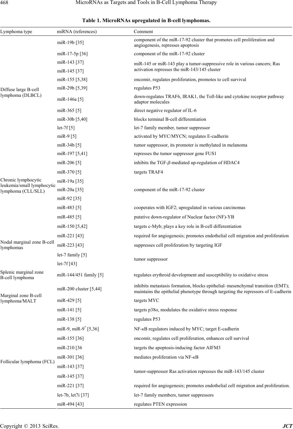 MicroRNAs as Targets and Tools in B-Cell Lymphoma Therapy Copyright © 2013 SciRes. JCT 468 Table 1. MicroRNAs upregulated in B-cell lymphomas. Lymphoma type miRNA (references) Comment miR-19b [35] component of the miR-17-92 cluster that promotes cell proliferation and angiogenesis, represses apoptosis miR-17-5p [36] component of the miR-17-92 cluster miR-143 [37] miR-145 [37] miR-145 or miR-143 play a tumor-suppressive role in various cancers; Ras activation represses the miR-143/145 cluster miR-155 [5,38] oncomir, regulates proliferation, promotes to cell survival miR-29b [5,39] regulates P53 miR-146a [5] down-regulates TRAF6, IRAK1, the Toll-like and cytokine receptor pathway adaptor molecules miR-365 [5] direct negative regulator of IL-6 miR-30b [5,40] blocks terminal B-cell differentiation let-7f [5] let-7 family member, tumor suppressor miR-9 [5] activated by MYC/MYCN; regulates E-cadherin Diffuse large B-cell lymphoma (DLBCL) miR-34b [5] tumor suppressor, its promoter is methylated in melanoma miR-197 [5,41] represses the tumor suppressor gene FUS1 miR-206 [5] inhibits the TGF-β-mediated up-regulation of HDAC4 miR-370 [5] targets TRAF4 miR-19a [35] miR-20a [35] miR-92 [35] component of the miR-17-92 cluster miR-483 [5] cooperates with IGF2; upregulated in various carcinomas Chronic lymphocytic leukemia/small lymphocytic lymphoma (CLL/SLL) miR-485 [5] putative down-regulator of Nuclear factor (NF)-YB miR-150 [5,42] targets c-Myb; plays a key role in B-cell differentiation miR-221 [43] required for angiogenesis; promotes endothelial cell migration and proliferation m Nodal marginal zone B-cell lymphomas iR-223 [43] suppresses cell proliferation by targeting IGF let-7 family [5] let-7f [43] tumor suppressor Splenic marginal zone B-cell lymphoma miR-144/451 family [5] regulates erythroid development and susceptibility to oxidative stress miR-200 cluster [5,44] inhibits metastasis formation, blocks epithelial–mesenchymal transition (EMT); maintains the epithelial phenotype through targeting the repressors of E-cadherin miR-429 [5] targets MYC Marginal zone B-cell lymphoma/MALT miR-141 [5] targets p38α, modulates the oxidative stress response miR-138 [5] regulates P53 miR-9, miR-9* [5,36] NF-κB regulators induced by MYC; target E-cadherin miR-155 [36] oncomir, regulates cell proliferation, enhances cell survival miR-210 [36 targets the apoptosis-inducing factor AIFM3 miR-301 [36] mediates proliferation via NF-κB miR-143 [37] miR-145 [37] tumor-suppressor Ras activation represses the miR-143/145 cluster miR-221 [37] required for angiogenesis; promotes endothelial cell migration and proliferation. let-7b, let7i [37] let-7 family members, tumor suppressors Follicular lymphoma (FCL) miR-494 [43] regulates PTEN expression 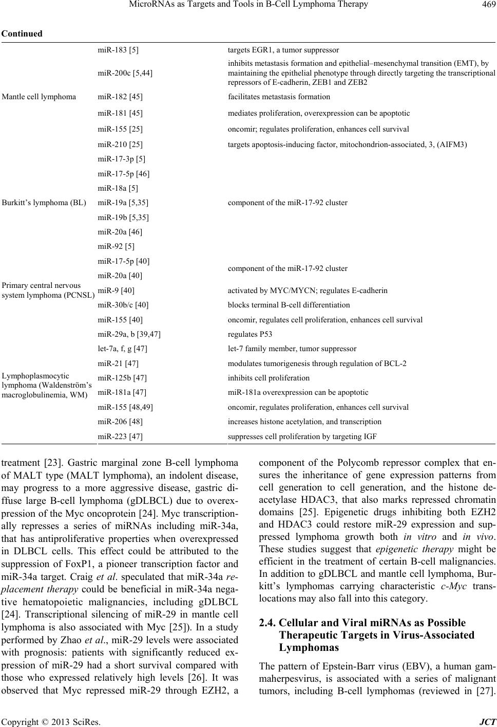 MicroRNAs as Targets and Tools in B-Cell Lymphoma Therapy 469 Continued miR-183 [5] targets EGR1, a tumor suppressor miR-200c [5,44] inhibits metastasis formation and epithelial–mesenchymal transition (EMT), by maintaining the epithelial phenotype through directly targeting the transcriptional repressors of E-cadherin, ZEB1 and ZEB2 miR-182 [45] facilitates metastasis formation miR-181 [45] mediates proliferation, overexpression can be apoptotic miR-155 [25] oncomir; regulates proliferation, enhances cell survival Mantle cell lymphoma miR-210 [25] targets apoptosis-inducing factor, mitochondrion-associated, 3, (AIFM3) miR-17-3p [5] miR-17-5p [46] miR-18a [5] miR-19a [5,35] miR-19b [5,35] miR-20a [46] Burkitt’s lymphoma (BL) miR-92 [5] component of the miR-17-92 cluster miR-17-5p [40] miR-20a [40] component of the miR-17-92 cluster miR-9 [40] activated by MYC/MYCN; regulates E-cadherin miR-30b/c [40] blocks terminal B-cell differentiation Primary central nervous system lymphoma (PCNSL) miR-155 [40] oncomir, regulates cell proliferation, enhances cell survival miR-29a, b [39,47] regulates P53 let-7a, f, g [47] let-7 family member, tumor suppressor miR-21 [47] modulates tumorigenesis through regulation of BCL-2 miR-125b [47] inhibits cell proliferation miR-181a [47] miR-181a overexpression can be apoptotic miR-155 [48,49] oncomir, regulates proliferation, enhances cell survival miR-206 [48] increases histone acetylation, and transcription Lymphoplasmocytic lymphoma (Waldenström’s macroglobulinemia, WM) miR-223 [47] suppresses cell proliferation by targeting IGF treatment [23]. Gastric marginal zone B-cell lymphoma of MALT type (MALT lymphoma), an indolent disease, may progress to a more aggressive disease, gastric di- ffuse large B-cell lymphoma (gDLBCL) due to overex- pression of the Myc oncoprotein [24]. Myc transcription- ally represses a series of miRNAs including miR-34a, that has antiproliferative properties when overexpressed in DLBCL cells. This effect could be attributed to the suppression of FoxP1, a pioneer transcription factor and miR-34a target. Craig et al. speculated that miR-34a re- placement therapy could be beneficial in miR-34a nega- tive hematopoietic malignancies, including gDLBCL [24]. Transcriptional silencing of miR-29 in mantle cell lymphoma is also associated with Myc [25]). In a study performed by Zhao et al., miR-29 levels were associated with prognosis: patients with significantly reduced ex- pression of miR-29 had a short survival compared with those who expressed relatively high levels [26]. It was observed that Myc repressed miR-29 through EZH2, a component of the Polycomb repressor complex that en- sures the inheritance of gene expression patterns from cell generation to cell generation, and the histone de- acetylase HDAC3, that also marks repressed chromatin domains [25]. Epigenetic drugs inhibiting both EZH2 and HDAC3 could restore miR-29 expression and sup- pressed lymphoma growth both in vitro and in vivo. These studies suggest that epigenetic therapy might be efficient in the treatment of certain B-cell malignancies. In addition to gDLBCL and mantle cell lymphoma, Bur- kitt’s lymphomas carrying characteristic c-Myc trans- locations may also fall into this category. 2.4. Cellular and Viral miRNAs as Possible Therapeutic Targets in Virus-Associated Lymphomas The pattern of Epstein-Barr virus (EBV), a human gam- maherpesvirus, is associated with a series of malignant tumors, including B-cell lymphomas (reviewed in [27]. Copyright © 2013 SciRes. JCT  MicroRNAs as Targets and Tools in B-Cell Lymphoma Therapy 470 Table 2. MicroRNAs down-regulated in B-cell lymphomas. Lymphoma type miRNA (references) Comments miR-145 [36] tumor suppressor in various cancers; Ras activation represses the miR-143/145 cluster miR-150 [36,42] regulates c-Myb; and plays a key role in B-cell differentiation miR-139 [36] suppresses metastasis and progression of liver carcinoma. miR-149 [36] miR-149* induces apoptosis by inhibiting Akt1 and E2F1 miR-320 [36] reduces ERK1/2 protein levels Diffuse large B-cell lymphoma (DLBCL) let-7e [36] tumor suppressor let-7 family (a, d, e, f, g) [5] let-7 family member, tumor suppressor effect miR-100 [5] suppresses IGF2 in breast cancer miR-125a [5] targets the negative NF-κB regulator TNFAIP3 miR-126, miR-126* [5,50] diagnostic for acute myeloid leukemia cases with common translocations miR-143 [5] tumor-suppressor; repressed by Ras activation. miR-146a [5] targets TRAF6 and IRAK1, the Toll-like and cytokine receptor pathway adaptor molecules miR-15b [51] miR-16 [51] miR-15 and miR-16 are direct transcriptional targets of E2F1 miR-17-5p [5] component of the miR-17-92cluster miR-20a,b [5] miR-182 [5] facilitates metastasis formation miR-223 [5] suppresses cell proliferation by targeting IGF miR-34a [5] tumor supressor miR-365 [5] direct negative regulator of IL-6 miR-451 [5] regulates erythroid development and susceptibility to oxidative stress miR-7 [5] inhibits tumor growth and metastasis miR-9, miR-9* [5] activated by MYC/MYCN, regulates E-cadherin Chronic lymphocytic leukemia/small lymphocytic lymphoma (CLL/SLL) miR-98 [5,41] targets the tumor suppressor FUS1 Nodal marginal zone B-cell lymphoma miR-370 [52] down-regulated in gastrointestinal tumors with 14q loss miR-141 [5,44] the miR-200 family inhibits the initiating step of metastasis, the epithelial mesenchymal transition (EMT), by maintaining the epithelial phenotype through directly targeting the transcriptional repressors of E-cadherin, ZEB1 and ZEB2 Splenic marginal zone B-cell lymphoma apoptosis-inducing factor, mitochondrion-associated, 3 (AIFM3) could be a direct target gene of miR-210 Marginal zone B-cell lymphoma/MALT miR-126* [5,50] diagnostic for acute myeloid leukemia cases with common translocations. miR-320 [36] reduces ERK1/2 protein levels and glucose induced ERK1/2 phosphorylation miR-149 [36] induces apoptosis by inhibiting Akt1 and E2F1 Follicular lymphoma (FCL) miR-139[36] suppresses metastasis and progression of hepatocellular carcinoma by down-regulating Rho-kinase 2. miR-126* [45,50] it’s expression distinguishes acute myeloid leukemia cases with common translocations miR-29a, b, c [25,39] regulates P53 miR-150 [25,42] regulates c-Myb expression; plays a key role in B-cell differentiation Mantle cell lymphoma miR-15a, b [25] miR-15 is a direct transcriptional targets of E2F1 cosing proliferation Copyright © 2013 SciRes. JCT  MicroRNAs as Targets and Tools in B-Cell Lymphoma Therapy 471 Continued let-7 family (a, d, e, f, g) [5] tumor suppressor miR-29 family a, b, c [5,39] regulates P53 miR-451 [5] regulates erythroid development, homeostasis and susceptibility to oxidative stress miR-146a [53] down-regulates TRAF6 and IRAK1, the Toll-like and cytokine receptor pathway adaptor molecules miR-150 [5,42] regulates c-Myb; plays a key role in B-cell differentiation Burkitt’s lymphoma (BL) miR-155 [53] oncogenic; regulates cell proliferation, enhances cell survival miR-145 [40] tumor-suppressor Primary central nervous system lymphoma (PCNSL) miR-214 [40] targets ATF4 miR-9 [47-49] activated by MYC/MYCN; regulates E-cadherin miR-152 [47] tumor suppressor miR-182 [47] facilitates metastasis formation miR-15a [48] tumor suppressor Lymphoplasmocytic lymphoma (Waldenström’s macroglobulinemia, WM) miR-16 [48] tumor suppressor One of the viral oncogenes expressed in nasopharyngeal carcinomas, post-transplant lymphomas, EBV-associated Hodgkin disease and in vitro transformed lymphoblastoid cell lines (LCLs) encodes a transmembrane protein called latent membrane protein 1 (LMP1) that affects the levels of certain cellular microRNAs by activating the NF-κB pathway. Cameron et al. observed that LMP1 induced miR-146a expression, down-regulating the products of a group of interferon-responsive genes [28]. miR-155, im- plicated in the development of B cell lymphomas, was also upregulated by LMP1 [29]. These virus-induced cel- lular miRNAs as well as the EBV-encoded miRNAs (re- viewed in [30]) are also potential targets of lymphoma therapy. 2.5. miRNAs as Potential Target Molecules in Radiotherapy and Chemotherapy of Mantle Cell Lymphoma Mantle cell lymphoma (MCL), comprising 5% - 10% of human B-cell malignancies is considered to be incurable with standard immuno-chemotherapy [31]. For this rea- son, a novel therapeutic strategy was elaborated, based on the delivery of radioisotopes to the tumor tissue using monoclonal antibodies. Anticipating the development of radioresistant MCL cells, Jiang et al. studied the effect of miRNA-17-92, an oncomir upregulated in various neo- plasms, on radiosensitivity of a human MCL cell line in vitro [32]. They observed that over-expression of mi- RNA-17-92 increased the survival of irradiated cells by indirectly up-regulating expression of the AKT protein kinase [32]. Thus, suppressing miRNA-17-92 may im- prove, in principle, the efficiency of MCL radiotherapy. It is worthy to note that another miRNA, miR-155, could be induced by hypoxia and protected lung cancer cells from radiation [33]. Treatment with anti-miR-155 mole- cules, however, radiosensitized the cells in vitro. Over- expression of miRNA-17-92 cluster inhibited chemothe- rapy-induced apoptosis in MCL cell lines in vitro [34]. miR-17-92 targeted a protein phosphatase involved in the negative regulation of the PI3K/AKT pathway, resulting in AKT activation. Rao et al. suggested that targeting the miRNA-17-92 cluster could be a plausible approach in MCL chemotherapy [34]. 3. Conclusion Recent developments in the synthesis of sophisticated, chemically modified antisense oligonucleotides targeting oncomirs and the use of anti-miR nanoparticle complexes made possible the development of new therapeutic ap- proaches that may complement current protocols for B- cell lymphoma therapy. Anti-cytokine therapy aiming at the removal of cytokines that activate the biogenesis of key oncomirs might also be considered in case of well defined patient groups. Suppression of oncomir expres- sion may successfully complement, on the long run, radio- therapy and chemotherapy of certain B-cell lymphoma types. In addition, switching on silenced tumor suppres- sor miRNAs by epigenetic drugs also appears to be a promising strategy in B-cell lymphoma treatment. 4. Acknowledgements We are grateful to Ms. Zsófia Papp for her help in the preparation of the manuscript. REFERENCES [1] D. Sayed and M. Abdellatif, “MicroRNAs in Develop- ment and Disease,” Physiological Reviews, Vol. 91, No. 3, 2011, pp. 827-887. doi:10.1152/physrev.00006.2010 [2] L. Ma and A. Weinberg, “MicroRNAs in Malignant Pro- Copyright © 2013 SciRes. JCT  MicroRNAs as Targets and Tools in B-Cell Lymphoma Therapy 472 gression,” Cell Cycle, Vol. 7, No. 5, 2008, pp. 570-572. doi:10.4161/cc.7.5.5547 [3] S. A. Melo and M. Esteller, “Dysregulation of microR- NAs in Cancer: Playing with Fire,” FEBS Letters, Vol. 585, No. 13, 2011, pp. 2087-2099. doi:10.1016/j.febslet.2010.08.009 [4] M. Fabbri and C. M. Croce, “Role of microRNAs in Lymphoid Biology and Disease,” Current Opinion in Hematology, Vol. 18, No. 4, 2011, pp. 266-272. doi:10.1097/MOH.0b013e3283476012 [5] L. Di Lisio, M. Sánchez-Beato, G. Gómez-López, M. E. Rodríguez, S. Montes-Moreno, M. Mollejo, J. Menárguez, M. A. Martínez, F. J. Alves, D. G. Pisano, M. A. Piris and N. Martínez, “MicroRNA Signatures in B-Cell Lympho- mas,” Blood Cancer Journal, Vol. 2, No. 2, 2012, Article ID: e57. doi:10.1038/bcj.2012.1 [6] P. S. Eis, W. Tam, L. Sun, A. Chadbun, Z. Li, M. F. Go- mez, E. Lund and J. E. Dahlberg, “Accumulation of miR- 155 and BIC RNA in Human B Cell Lymphomas,” Pro- ceedings of the National Academy of Sciences, USA, Vol. 102, No. 10, 2005, pp. 3627-3632. doi:10.1073/pnas.0500613102 [7] J. Kluiver, S. Poppema, D. de Jong, T. Blokzijl, G. Harms, S. Jacobs, B. J. Kroessen and A. van den Berg, “BIC and miR-155 Are Highly Expressed in Hodgkin, Primary Me- diastinal and Diffuse Large B Cell Lymphomas,” Journal of Pathology, Vol. 207, No. 2, 2005, pp. 243-249. doi:10.1002/path.1825 [8] S. Costinean, N. Zanesi, Y. Pekarsky, E. Tili, S. Volinia, N. Heerema and C. M. Croce, “Pre-B Cell Proliferation and Lymphoblastic Leukemia/High-Grade Lymphoma in E(mu)-miR155 Transgenic Mice,” Proceedings of the Na- tional Academy of Sciences, USA, Vol. 103, No. 18, 2006, pp. 7024-7029. doi:10.1073/pnas.0602266103 [9] S. Costinean, S. K. Sandhu, I. M. Pedersen, E. Tili, R. Trotta, D. Perrotti, D. Ciarlariello, P. Neviani, J. Harb, L. R. Kauffmann, A. Shidham and C. M. Croce, “Src Ho- mology Domain-Containing Inositol-5-Phosphatase and CCAAT Enhancer-Binding Protein β Are Targeted by miR-155 in B Cells of Eμ-MIR-155 Transgenic Mice,” Blood, Vol. 114, No. 7, 2009, pp. 1374-1382. doi:10.1182/blood-2009-05-220814 [10] C. L. Li, H. Nie, M. Wang, L. P. Su, J. F. Li, Y. Y. Yu, M. Yan, Q. L. Qu, Z. G. Zhu and B. Y. Liu, “microRNA-155 Is Downregulated in Gastric Cancer Cells and Involved in Cell Metastasis,” Oncology Report, Vo. 27, No. 6, 2012, pp. 1960-1966. doi:10.3892/or.2012.1719 [11] I. A. Babar, C. J. Cheng, C. J. Booth, X. Liang, J. B. Weidhaas, W. M. Saltzman and F. J. Slack, “Nanoparti- cle-based Therapy in an in Vivo microRNA-155 (miR- 155)-Dependent Mouse Model of Lymphoma,” Proceed- ings of the National Academy of Sciences, USA, Vol. 109, No. 26, 2012, pp. E1695-E1704. doi:10.1073/pnas.1201516109 [12] A. G. Torres, M. M. Fabiani, E. Vigorito and M. J. Gait, “MicroRNA Fate upon Targeting with Anti-miRNA Oli- gonucleotides as Revealed by an Improved Northern- Blot-Based Method for miRNA Detection,” RNA-A Pub- lication of RNA Society, Vol. 17, No. 5, 2011, pp. 933- 943. doi:10.1261/rna.2533811 [13] O. Snove and J. J. Rossi, “Toxicity in Mice Expressing Short Hairpin RNAs Gives New Insight into RNAi,” Ge- nome Biology, Vol. 7, No. 8, 2006, Article ID: 231. doi:10.1186/gb-2006-7-8-231 [14] D. Peer and M. Shimaoka, “Systemic siRNA Delivery to Leukocyte-Implicated Diseases,” Cell Cycle, Vol. 8, No. 6, 2009, pp. 853-859. doi:10.4161/cc.8.6.7936 [15] M. M. Fabani and M. J. Gait, “miR-122 Targeting with LNA/2’-O-Methyl Oligonucleotide Mixmers, Peptide Nu- cleic Acids (PNA), and PNA-Peptide Conjugates,” RNA- A Publication of RNA Society, Vol. 14, No. 2, 2008, pp. 336-346. doi:10.1261/rna.844108 [16] M. M. Fabani, C. Abreu-Goodger, D. Williams, P. A. Lyons, A. G. Torres, K. G. C. Smith, A. J. Enright, M. J. Gait and E. Vigorito, “Efficient Inhibition of miR-155 Function in Vivo by Peptide Nucleic Acids,” Nucleic Ac- ids Research, Vol. 38, No. 13, 2010, pp. 4466-4475. doi:10.1093/nar/gkq160 [17] J. Krützfeldt, S. Kuwajima, R. Braich, K. G. Rajeev, J. Pena, T. Tuschl, M. Manoharan and M. Stoffel, “Speci- ficity, Duplex Degradation and Subcellular Localization of Antagomirs,” Nucleic Acids Research, Vol. 35, No. 9, 2007, pp. 2885-2892. doi:10.1093/nar/gkm024 [18] Y. Zhang, A. M. Roccaro, C. Rombaoa, L. Flores, S. Obad, S. M. Fernandes, A. Sacco, Y. Liu, H. Ngo, P. Quang, A. K. Azab, F. Azab, P. Maiso, M. Reagan, J. R. Brown, T. H. Thai, S. Kauppinen and I. M. Ghobrial, “LNA-Mediated Anti-miR-155-Silencing in Low Grade B-Cell Lymphomas,” Blood, Vol. 120, No. 8, 2012, pp. 1678-1686. doi:10.1182/blood-2012-02-410647 [19] V. Prabhu, S. Uzzaman, V. M. B. Grace and C. Guru- vayoorappan, “Nanoparticles in Drug Delivery and Can- cer Therapy: The Giant Rats Tail,” Journal of Cancer Therapy, Vol. 2, 2011, pp. 325-334. doi:10.4236/jct.2011.23045 [20] J. Su, H. Baigude, J. McCarroll and T. M. Rana, “Silenc- ing microRNA by Interfering Nanoparticles in Mice,” Nucleic Acids Research, Vol. 39, No. 6, 2011, Article ID: e38. doi:10.1093/nar/gkq1307 [21] T. H. Thai, D. P. Calado, K. M. Ansel, C. Xiao, Y. Xue, A. Murphy, D. Frendewey, D. Valenzuela, J. L. Kutok, M. Schmidt-Supprian, N. Rajewsky, G. Yancopoulos, A. Rao and K. Rajewsky, “Regulation of the Germinal Center Response by microRNA-155,” Science, Vol. 316, No. 5824, 2007, pp. 604-608. doi:10.1126/science.1141229 [22] I. Pedersen, D. Otero, A. V. Miletic, C. Hother, E. Ralfki- aer, R. C. Rickert, K. Gronbaek and M. David, “Onco- miR-155 Targets SHIP1 to Promote TNFα-Dependent Growth of B Cell Lymphomas,” EMBO Molecular Medi- cine, Vol. 1, No. 5, 2009, pp. 288-295. doi:10.1002/emmm.200900028 [23] Y. A. Mian and N. J. Zeleznik-Le, “MicroRNAs in Leu- kemias: Emerging Diagnostic Tools and Therapeutic Tar- gets,” Current Drug Targets, Vol. 11, No. 7, 2010, pp. 801-811. doi:10.2174/138945010791320872 [24] V. J. Craig, S. B. Cogliatti, J. Imig, C. Renner, S. Neuen- schwander, H. Rehrauer, R. Schlapbach, S. Dimhofer, A. Copyright © 2013 SciRes. JCT  MicroRNAs as Targets and Tools in B-Cell Lymphoma Therapy 473 Tzankov and A. Müller, “Myc-Mediated Repression of microRNA-34a Promotes High-Grade Transformation of B Cell Lymphoma by Dysregulation of FoxP1,” Blood, Vol. 117, No. 23, 2011, pp. 6227-6236. [25] X. Zhang, X. Zhao, W. Fiskus, J. Lin, T. Lwin, R. Rao, Y. Zhang, J. C. Chan, K. Fu, V. E. Marquez, S. Chen-Kiang, L. C. Moscinski, E. Seto, W. S. Dalton, K. L. Wright, E. Sotomayor, K. Bhalla and J. Tao, “Coordinated Silencing of MYC-Mediated miR-29 by HDAC3 and EZH2 as a Therapeutic Target of Histone Modification in Aggressive B Cell Lymphoma,” Cancer Cell, Vol. 22, No. 4, 2012, pp. 506-523. doi:10.1016/j.ccr.2012.09.003 [26] J. J. Zhao, J. Lin, T. Lwin, H. Yang, J. Guo, W. Kong, S. Dessureault, L. C. Moscinski, D. Rezania, W. S. Dalton, E. Sotomayor, J. Tao and J. Q. Cheng, “microRNA Ex- pression Profile and Identification of miR-29 as a Prog- nostic Marker and Pathogenic Factor by Targeting CDK6 in Mantle Cell Lymphoma,” Blood, Vol. 115, No. 13, 2010, pp. 2630-2639. doi:10.1182/blood-2009-09-243147 [27] M. Takacs, F. Banati, A. Koroknai, J. Segesdi, D. Sala- mon, H. Wolf, H. H. Niller and J. Minarovits, “Epigenetic Regulation of Latent Epstein-Barr Virus Promoters,” Bio- chimica et Biophysica Acta, Vol. 1799, No. 3-4, 2010, pp. 228-235. doi:10.1016/j.bbagrm.2009.10.005 [28] J. E. Cameron, Q. Yin, C. Fewell, M. Lacey, J. McBride, X. Wang, Z. Lin, B. C. Schaefer and E. K. Flemington, “Epstein-Barr Virus Latent Membrane Protein 1 Induces Cellular microRNA miR-146A, a Modulator of Lympho- cyte Signaling Pathways,” Journal of Virology, Vol. 82, No. 4, 2008, pp. 1946-1958. doi:10.1128/JVI.02136-07 [29] F. Lu, A. Weidmer, C. G. Liu, S. Volnia, C. M. Croce and P. M. Lieberman, “Epstein-Barr Virus-Induced miR-155 Attenuates NF-kappaB Signaling and Stabilizes Latent Virus Persistence,” Journal of Virology, Vol. 82, No. 21, 2008, pp. 10436-10443. doi:10.1128/JVI.00752-08 [30] B. R. Cullen, “Viruses and microRNAs: RISCy Interac- tions with Serious Consequences,” Genes & Development, Vol. 25, No. 18, 2011, pp. 1881-1894. [31] A. P. Skarbnik and M. R. Smith, “Radioimmunotherapy in Mantle Cell Lymphoma,” Best Practice & Research Clinical Haematology, Vol. 25, No. 2, 2012, pp. 201-210. doi:10.1016/j.beha.2012.04.004 [32] P. Jiang, E. Y. Rao, N. Meng, Y. Zhao and J. J. Wang, “MicroRNA-17-92 Significantly Enhances Radioresis- tance in Human Mantle Cell Lymphoma Cells,” Radia- tion Oncology, Vol. 5, 2010, Article ID: 100. doi:10.1186/1748-717X-5-100 [33] I. A. Babar, J. Czochor, A. Steinmetz, J. B. Weidhaas, P. M. Glazer and F. J. Slack, “Inhibition of Hypoxia-In- duced miR-155 Radiosensitizes Hypoxic Lung Cancer Cells,” Cancer Biology & Therapy, Vol. 12, No. 10, 2012, pp. 908-914. doi:10.4161/cbt.12.10.17681 [34] E. Rao, C. Jiang, M. Ji, X. Huang, J. Ibgal, G. Lenz, G. Wright, L. M. Staudt, Y. Zhao, T. W. McKeithan, W. C. Chan and K. Fu, “The miRNA-17~92 Cluster Mediates Chemoresistance and Enhances Tumor Growth in Mantle Cell Lymphoma via PI3K/AKT Pathway Activation,” Leukemia, Vol. 26, No. 5, 2012, pp. 1064-1072. doi:10.1038/leu.2011.305 [35] J. H. Gibcus, L. P. Tan, G. Harms, R. N. Schakel, D. de Jong, T. Blokzijl, P. Möller, S. Poppema, B. J. Kroesen and A. van den Berg, “Hodgkin Lymphoma Cell Lines Are Characterized by a Specific miRNA Expression Pro- file,” Neoplasia, Vol. 11, No. 2, 2009, pp. 167-176. [36] A. Roehle, K. P. Hoefig, D. Repsilber, C. Thorns, M. Ziepert, K. O. Wesche, M. Thiere, M. Loeffler, W. Klap- per, M. Pfreundschuh, A. Matolcsy, H. W. Bernd, L. Re- iniger, H. Merz and A. C. Feller, “MicroRNA Signatures Characterize Diffuse Large B-Cell Lymphomas and Fol- licular Lymphomas,” British Journal of Haematology, Vol. 142, No. 5, 2008, pp. 732-744. doi:10.1111/j.1365-2141.2008.07237.x [37] C. H. Lawrie, J. Chi, S. Taylor, D. Tramonti, E. Ballabio, S. Palazzo, N. J. Saunders, F. Pezzella, J. Boultwood, J. S. Wainscoat and C. S. Hatton, “Expression of microRNAs in Diffuse Large B Cell Lymphoma Is Associated with Immunophenotype, Survival and Transformation from Follicular Lymphoma,” Journal of Cellular and Mole- cular Medicine, Vol. 13, No. 7, 2009, pp. 1248-1260. doi:10.1111/j.1582-4934.2008.00628.x [38] D. Rai, S. Karanti, I. Jung, P. L. Dahia and R. C. Aguiar, “Coordinated Expression of microRNA-155 and Pre- dicted Target Genes in Diffuse Large B-Cell Lym- phoma,” Cancer Genetics and Cytogenetics, Vol. 181, No. 1, 2008, pp. 8-15. doi:10.1016/j.cancergencyto.2007.10.008 [39] S. Y. Park, J. H. Lee, M. Ha, J. W. Nam and V. N. Kim, “miR-29 miRNAs Activate p53 by Targeting p85 Alpha and CDC42,” Nature Structural and Molecular Biology, Vol. 16, No. 1, 2009, pp. 23-29. doi:10.1038/nsmb.1533 [40] L. Fischer, M. Hummel, A. Korfel, D. Lenze, K. Joehrens and E. Thiel, “Differential micro-RNA Expression in Pri- mary CNS and Nodal Diffuse Large B-Cell Lympho- mas,” Neuro-Oncology, Vol. 13, No. 10, 2011, pp. 1090- 1098. doi:10.1093/neuonc/nor107 [41] L. Du, J. J. Schageman, M. C. Subauste, B. Saber, S. M. Hammond and L. Prudkin, “miR-93, miR-98, and miR- 197 Regulate Expression of Tumor Suppressor Gene FUS1,” Molecular Cancer Research, Vol. 7, No. 8, 2009, pp. 1234-1243. doi:10.1158/1541-7786.MCR-08-0507 [42] C. Xiao, D. P. Calado, G. Galler, T. H. Thai, H. C. Pat- terson, J. Wang, N. Rajewsky, T. P. Bender and K. Ra- jewsky, “MiR-150 Controls B Cell Differentiation by Targeting the Transcription Factor c-Myb,” Cell, Vol. 131, No. 1, 2007, pp. 146-159. doi:10.1016/j.cell.2007.07.021 [43] A. J. Arribas, Y. Campos-Martín, C. Gómez-Abad, P. Al- gara, M. Sánchez-Beato, M. S. Rodriguez-Pinilla, S. Mon- tes-Moreno, N. Martinez, J. Alves-Ferreira, M. A. Piris and M. Mollejo, “Nodal Marginal Zone Lymphoma: Gene Expression and miRNA Profiling Identify Diagnos- tic Markers and Potential Therapeutic Targets,” Blood, Vol. 119, No. 3, 2012, pp. e9-e21. doi:10.1182/blood-2011-02-339556 [44] S. M. Park, A. B. Gaur, E. Lengyel and M. E. Peter, “The miR-200 Family Determines the Epithelial Phenotype of Cancer Cells by Targeting the E-Cadherin Repressors ZEB1 and ZEB2,” Genes Development, Vol. 22, No. 7, 2008, pp. 894-907. doi:10.1101/gad.1640608 Copyright © 2013 SciRes. JCT  MicroRNAs as Targets and Tools in B-Cell Lymphoma Therapy Copyright © 2013 SciRes. JCT 474 [45] L. Di Lisio, G. Gomez-Lopez, M. Sanchez-Beato, C. Gomez-Abad, M. E. Rodriguez, R. Villuendas, B. I. Fer- reira, A. Carro, D. Rico, M. Mollejo, M. A. Martínez, J. Menárguez, A. Díaz-Alderete, J. Gil, J. C. Cigudosa, D. G. Pisano, M. A. Piris and N. Martínez, “Mantle Cell Lymphoma: Transcriptional Regulation by microRNAs,” Leukemia, Vol. 24, No. 7, 2010, pp. 1335-1342. doi:10.1038/leu.2010.91 [46] A. Onnis, G. de Falco, G. Antonicelli, M. Onorati, C. Bellan, O. Sherman, S. Sayed and L. Leoncini, “Altera- tion of microRNAs Regulated by c-Myc in Burkitt Lym- phoma,” PLoS ONE, Vol. 5, No. 9, 2010, p. e12960. doi:10.1371/journal.pone.0012960 [47] L. S. Hodge, S. F. Elsawa, D. M. Grote, T. L. Price- Troska, Y. W. Asmann, R. Fonseca, M. A. Gertz, T. E. Witzig, A. J. Novak and S. M. Ansell, “MicroRNA Ex- pression in Tumor Cells from Waldenstrom’s Macro- globulinemia Reflects Both Their Normal and Malignant Cell Counterparts,” Blood Cancer Journal, Vol. 1, No. 6, 2011, Article ID: e24. doi:10.1038/bcj.2011.25 [48] E. Braggio, C. Philipsborn, A. Novak, L. Hodge, S. An- sell and R. Fonseca, “Molecular Pathogenesis of Walden- strom’s Macroglobulinemia,” Haematologica, Vol. 97, No. 9, 2012, pp. 1281-1290. doi:10.3324/haematol.2012.068478 [49] A. M. Roccaro, A. Sacco, C. Chen, J. Runnels, X. Leleu, F. Azab, A. K. Azab, X. Jia, H. T. Ngo, M. R. Melhem, N. Burwick, L. Varticovski, C. D. Novina, B. J. Rollins, K. C. Anderson and I. M. Ghobrial, “MicroRNA Expression in the Biology, Prognosis, and Therapy of Waldenström Macroglobulinemia,” Blood, Vol. 113, No. 18, 2009, pp. 4391-4402. doi:10.1182/blood-2008-09-178228 [50] Z. Li, J. Lu, M. Sun, S. Mi, H. Zhang, R. T. Luo, P. Chen, Y. Wang, M. Yan, Z. Qian, M. B. Neilly, J. Jin, Y. Zhang, S. K. Bohlander, D. E. Zhang, R. A. Larson, M. M. Le Beau, M. J. Thirman, T. R. Golub, J. D. Rowley and J. Chen, “Distinct microRNA Expression Profiles in Acute Myeloid Leukemia with Common Translocations,” Pro- ceedings of the National Academy of Sciences of the United States of America, Vol. 105, No. 40, 2008, pp. 15535-15540. doi:10.1073/pnas.0808266105 [51] G. A. Calin, C. D. Dumitru, M. Shimizu, R. Bichi, S. Zupo, E. Noch, H. Aldler, S. Rattan, M. Keating K. Rai, L. Rassenti, T. Kipps, M. Negrini, F. Bullrich and C. M. Croce, “Frequent Deletions and Down-Regulation of Micro-RNA Genes miR15 and miR16 at 13q14 in Chro- nic Lymphocytic Leukemia,” Proceedings of the National Academy of Sciences of the United States of America, Vol. 99, No. 24, 2002, pp. 15524-15529. doi:10.1073/pnas.242606799 [52] F. Haller, A. Von Heydebreck, J. D. Zhang, B. Gunawan, C. Langer, G. Ramadori, S. Wiemann and O. Sahin, “Lo- calization- and Mutation-Dependent microRNA (miRNA) Expression Signatures in Gastrointestinal Stromal Tu- mours (GISTs), with a Cluster of Co-Expressed miRNAs Located at 14q32.31,” Journal of Pathology and Bacteri- ology, Vol. 220, No. 1, 2010, pp. 71-86. doi:10.1002/path.2610 [53] J. L. Robertus, J. Kluiver, C. Weggemans, G. Harms, R. M. Reijmers, Y. Swart, K. Kok, S. Rosati, E. Schuuring, G. van Imhoff, S. T. Pals, P. Kluin and A. van den Berg, “MiRNA Profiling in B Non-Hodgkin Lymphoma: A MYC-Related miRNA Profile Characterizes Burkitt Lym- phoma,” British Journal of Haematology, Vol. 149, No. 6, 2010, pp. 896-899. doi:10.1111/j.1365-2141.2010.08111.x
|