Open Journal of Stomatology, 2013, 3, 504-506 OJST
http://dx.doi.org/10.4236/ojst.2013.39082 Published Online December 2013 (http://www.scirp.org/journal/ojst/)
Calcifying cystic odontogenic tumour manifesting in the
maxillary antrum: Case report
Eunice Kihara1, Richard Owino2, Josiah Otwoma1, Elizabeth Dimba1, Mark Chindia1
1Department of Oral & Maxillofacial Surgery—Oral Pathology & Oral Medicine, University of Nairobi, Nairobi, Kenya
2Department of Paediatric Dentistry & Orthodontics, University of Nairobi, Nairobi, Kenya
Email: eunik20@yahoo.com
Received 31 October 2013; revised 1 December 2013; accepted 12 December 2013
Copyright © 2013 Eunice Kihara et al. This is an open access article distributed under the Creative Commons Attribution License,
which permits unrestricted use, distribution, and reproduction in any medium, provided the original work is properly cited.
ABSTRACT
A case is presented of a 15-year-old boy who mani-
fested the calcifying cystic odontogenic tumour (CCOT)
in the left maxillary antrum of an unknown duration.
In addition, the patient had a high arched palate and
multiple impacted teeth of the normal series and su-
pernumerary type including mesiodens as demon-
strated in an orthopantomograph.
Keywords: Case Reports; Odontogenic Cyst; Calcifying;
Odontogenic Tumours; Maxillary Sinus
1. INTRODUCTION
Generally, benign odontogenic neoplasms are relatively
rare lesions that are derived from either the epithelial an d
mesenchymal components or the remnant elements of the
developing tooth germs. These lesions are often classi-
fied according to location as p eripheral or central entities
[1-4]. In this category of diseases, the calcifying cystic
odontogenic tumour (CCOT) is a particularly rare benign
entity that is characterized by an ameloblastoma-like
epithelium and ghost cells that have the potential to un-
dergo calcification [4-6]. The lesion usually appears as a
painless, slow-growing tumour that may involve the
maxilla or mandible in the anterior segments [1,3]. We
report a 15-year-old boy who presented with CCOT man-
ifesting in the left maxillary antrum associated with den-
tal malformation, tooth impaction and a high arched pal-
ate.
2. CASE REPORT
A 15-year-old boy was referred to our clinic for evalua-
tion and management of a persistent swelling of the left
maxilla that had, apparently, been noted nearly two years
previously. Over that period, the swelling had been es-
sentially painless. However, the patient reported that the
swelling had then become painful. In addition, he also
experienced a feeling of stuffiness in the left nostril. The
patient’s general medical and dental history was unre-
markable. On examination, the patient manifested a non-
tender mass causing facial asymmetry with fullness in
the left nasolabial fo ld extending towards the bod y of the
zygoma. Intraorally, there was total left maxillary sulcu-
lar fullness, a mixed dentition and a generalized malposi-
tioning of some teeth in both arches in addition to a high
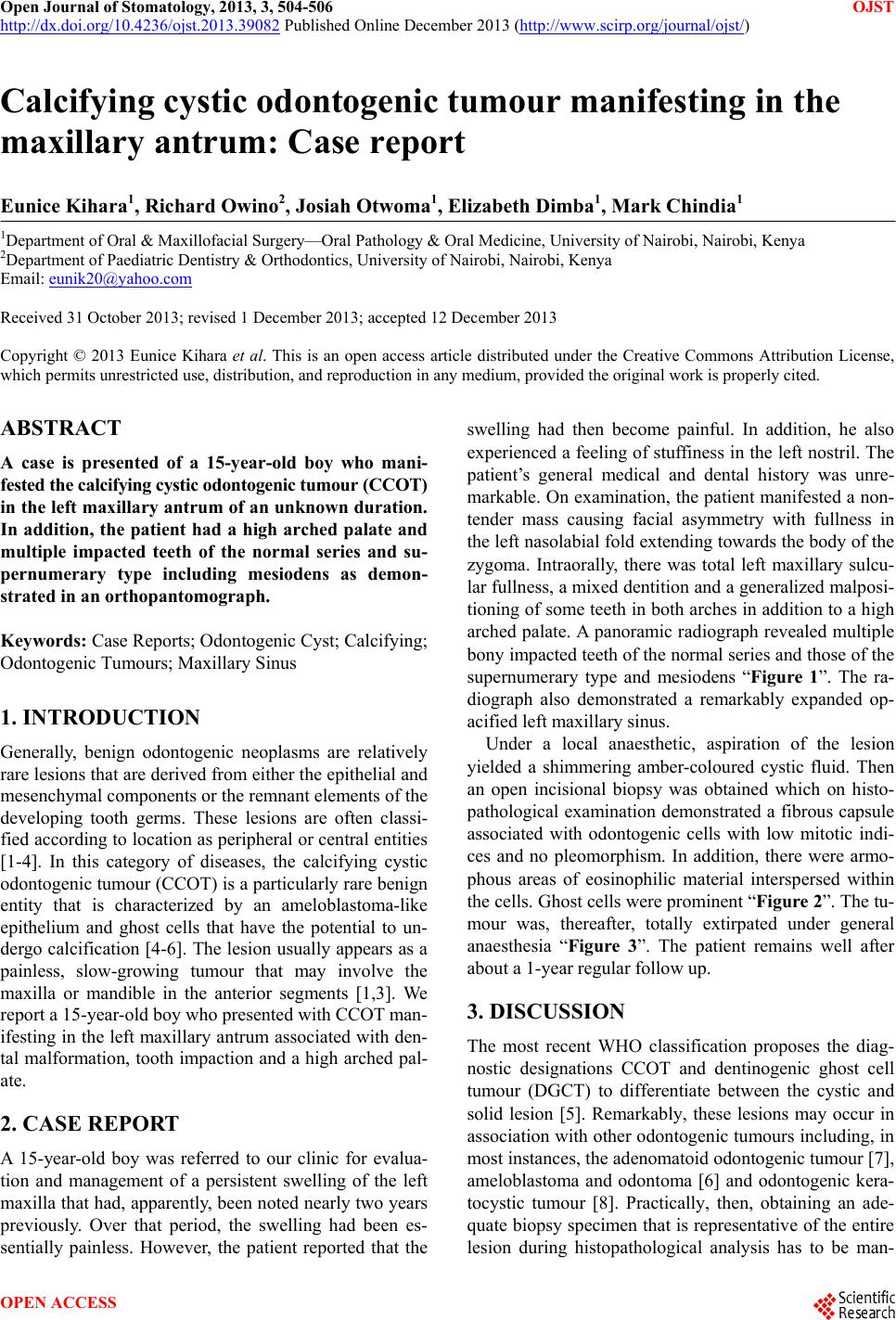
arched palate. A panoramic radiograph rev ealed multiple
bony impacted teeth of the normal series and thos e of the
supernumerary type and mesiodens “Figure 1”. The ra-
diograph also demonstrated a remarkably expanded op-
acified left maxillary sinus.
Under a local anaesthetic, aspiration of the lesion
yielded a shimmering amber-coloured cystic fluid. Then
an open incisional biopsy was obtained which on histo-
pathological examination demonstrated a fibrous capsule
associated with odontogenic cells with low mitotic indi-
ces and no pleomorphism. In addition, there were armo-
phous areas of eosinophilic material interspersed within
the cells. Ghost cells were prominent “Figure 2”. The tu-
mour was, thereafter, totally extirpated under general
anaesthesia “Figure 3”. The patient remains well after
about a 1-year regular follow up.
3. DISCUSSION
The most recent WHO classification proposes the diag-
nostic designations CCOT and dentinogenic ghost cell
tumour (DGCT) to differentiate between the cystic and
solid lesion [5]. Remarkably, these lesions may occur in
association with other odontogenic tumours including, in
most instances, the adenomatoid odontogenic tumour [7],
ameloblastoma and odontoma [6] and odontogenic kera-
tocystic tumour [8]. Practically, then, obtaining an ade-
quate biopsy sp ecimen that is represen tative of the entire
lesion during histopathological analysis has to be man-
OPEN ACCESS

