1. Introduction
All present dosimetry protocols [1] [2] [3] [4] recommend the use of well- guarded, air-filled parallel-plate ion chambers for reference dosimetry in clinical electron beams. The recommendation of well-guarded chambers is especially based on report 35 of the International Commission on Radiation Units & Measurements (ICRU) [5] which was the principles of clinical electron dosimetry that are summarized. In this report, the so-called in-scattering effect is described in detail: based on the strong reduction of the energy losses and multiple scattering of the electrons in the gas-filled cavity compared to the surrounding phantom material, more electrons are scattered into than out of the cavity. As a result, at the lateral boundary of the air-filled cavity, a dose oscillation arises (see Figure (4.2) in ICRU 35) resulting in an over-response of the air-filled cavity, which according to all dosimetry protocols, has to be corrected by a fluence perturbation correction pcav. In attempt to make the chamber signal insensitive to the in/out electrontransport imbalance and thereby, bringing pcav to unity, modern parallel-plate chambers are equipped with a wide guard ring to keep the region of fluence perturbation at a safe distance from the chamber’s collecting volume.
Moreover, all present dosimetry protocols assume a negligible influence of the entrance window and the surrounding wall material on the response of modern parallel-plate chambers, i.e. the wall perturbation correction defined in all dosimetry protocols is assumed to be unity.
In a previous publication, Zink et al. [6] reinvestigated in detail the in- and out-scattering of electrons in gas-filled cavities, which gave a new insight into the perturbation correction pcav for parallel-plate chambers in clinical electron beams. With the help of spatially resolved Monte Carlo calculations, they have shown that the in-scattering effect indeed exists, but they have also shown that a guard ring has only a minor effect on the dose to a gas-filled cavity, especially for cavities with small diameters as in the case of the Markus chamber. The cavity diameter itself has a much larger impact on the dose within the cavity. This is a consequence of the deep radial penetration of the in- and out-bound transport of electrons into the gas-filled cavity. These results question not only the relevance of the guard ring for this chamber type but also the value of the perturbation correction pcav for the guardless Markus chamber given in all present dosimetry protocols. These values are all based on an experimental study performed by Van der Pleatsen et al. [7] in the early 1990s. They compared the chamber’s dose D for the guardless Markus chamber with the dose of a NACP chamber in clinical electron beams, assuming that the NACP chamber represents a perturbation-free ion chamber. The ratio  was interpreted as the fluence perturbation correction pcav for the Markus chamber.
The aim of the present study is a Monte Carlo based reiteration of Van der Plaetsen’s experiment against the background of the actual knowledge about the in-scattering effect in gas-filled cavities in clinical electron beams. The present data may be especially important regarding the planned update of the international dosimetry protocol IAEA TRS-398 [2] .
2. Fundamentals
Following Bragg-Gray theory, the absorbed dose to water DW may be derived from the dose to the air-filled detector Ddet, the restricted stopping power ratio  of the materials water w and air a, and a perturbation correction p [5] [8] :
 (1)
(1)
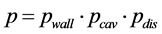
It is assumed that the perturbation factor p may be factorized, for parallel- plate chambers it is traditionally split into three components:
 (2)
(2)
where pwall stands for the fluence perturbation due to the chamber wall, pcav the in-scattering of electrons from the surrounding phantom material into the air- filled cavity (Pfl in The American Association of Physicists in Medicine (AAPM) TG-51 [1] ), and pdis for fluence changes due to the replacement of the phantom material water by the air-filled cavity (Pgr in AAPM TG-51).
For parallel-plate chambers pdis equals unity according to all present dosimetry protocols when the reference point of the chamber (center of the entrance plane of the air-filled cavity) is positioned at the depth of measurement z. Because of the thin entrance window of all parallel-plate chamber types, also the wall perturbation pwall is assumed to be unity. As the NACP chamber is equipped with a wide guard ring (w = 0.33 cm), Van der Plaetsen et al. [7] moreover assumed that for this chamber type also pcav is unity for all electron energies, i.e. the NACP chamber was considered a perturbation-free ion chamber. Therefore, the dose ratio  is interpreted as the perturbation correction pcavfor the Markus chamber due to in-scattering electrons as this chamber type is not equipped with a guard ring of sufficient width (w = 0.035 cm). The dose ratio was determined for several primary electron energies at the depth of the dose maximum zmax and could be fitted by a function of the mean electron energy
  at this depth:
 (3)
(3)
In the IAEA protocol [2] , this equation was adapted to the actual beam quality specifier R50 and the reference depth zref:
 (4)
(4)
3. Methods and Material
Comparable to the experiments conducted by Van der Plaetsen, the dose ratio was calculated for the NACP chamber and additionally for the other well-guarded chambers, the Roos and the Advanced Markus chambers, using the Monte Carlo code EGSnrc (V4 2.4.0) [9] [10] [11] [12] . The ion chambers were modeled in detail with the egs++ geometry package according to the blueprints provided by the manufacturer PTW [13] . In the case of the NACP-02 chamber, the geometry is based on the information given in several publications [14] - [19] . Geometric details of the chambers with their material components are summarized in Table 1 and Figure 1. The investigation was performed with thirteen clinical electron spectra (6 MeV < E0 < 21 MeV) taken from literature [20] and a full modeled Elekta Synergy accelerator including an electron applicator with a field size of 10 × 10 cm2 (see Table 2 for details). For the accelerator model the energies of the primary electrons hitting the scattering foil were E0 = 6, 12 and 18 MeV. The accelerator was modeled with the BEAMnrc code [21] according to the blueprints provided by the manufacturer.
The user code egs_chamber [22] was applied for the calculation of the dose
![]()
Table 1. Geometric details of the modeled parallel-plate chambers. V is the active chamber volume, r the radius of the active volume, h its height and w the width of the guard ring. Additionally the entrance window thickness d is given.
![]()
Figure 1. Schematic illustration of the outer dimension and materials of the used parallel-plate chambers: (a) Roos, (b) NACP, (c) Adv. Markus and (d) Markus. The green rectangle represents the air-filled cavity for all chambers. For the Roos, Markus and Adv. Markus the outer material PMMA is given in red. In contrast for die NCAP the outer material polystyrene is drafted in blue. The Markus and Adv. Markus chambers have a small air gap above their sensitive cavities. The NACP chamber has inside parts of 1.82 g/cm3 carbon given in claret.
![]()
Table 2. Characteristic data of the electron sources applied in this study. The Elekta Synergy accelerator was modeled in detail including the electron applicator, for the other accelerators only spectra were used as electron sources [19] . The given data are the mean electron energy at the depth of the dose maximum  and at the reference depth, the corresponding depths zmax and zref and the electron beam specifier R50.
deposition  within the detectors and within a small water voxel (r = 0.5 cm, z = 0.02 cm) to determine the dose to water DW.
To enable a comparison of the Monte Carlo data with the original data from Van der Plaetsen and with the data given in the IAEA protocol, the simulations were performed for two depths within a water phantom (30 × 30 × 30 cm3): the depth of the dose maximum zmax and the reference depth zref. In all cases, the chambers were positioned with their reference point at the correspondent depth. The source-to-surface distance was 100 cm and the field size at the phantom surface 10 × 10 cm2. Also to enable comparability with Van der Plaetsen we additionally determined the mean electron energy  at the depth of measurement. The determination of the mean electron energies at depth z within the water phantom was performed with the user code FLURZnrc [23] . To calculate the total perturbation correction p the dose to water was also calculated at depths zmax and zref within a small water voxel. To avoid the calculation of the stopping power ratios
 , the cavities of the chambers were filled with low- density water, i.e. water with the density of air, and a density correction corresponding to normal-density water [24] . In that case, the perturbation correction p can simply derived from the dose ratio
 , i.e.
 . The cutoff/threshold energies for the particle transport were set to 512 keV for electrons and 10 keV for photons; all other EGS parameters were set to their default values.
4. Results
Figure 2 shows the ratio of the dose to the active volume of the well-guarded Roos, NACP and Advanced Markus chambers to the dose within the guardless Markus chamber. In the upper panel this dose ratio is given for the depth of the dose maximum zmax as a function of the mean electron energy![]() , i.e. these data are directly comparable with the results published by Van der Plaetsen. The fit according to Equation (3) is additionally shown. As can be seen, the dose for all guarded chambers is quite similar; for all energies they do not deviate from each other by more than 0.3%. For the largest mean energy
, i.e. these data are directly comparable with the results published by Van der Plaetsen. The fit according to Equation (3) is additionally shown. As can be seen, the dose for all guarded chambers is quite similar; for all energies they do not deviate from each other by more than 0.3%. For the largest mean energy![]() , corresponding
, corresponding
![]()
Figure 2. Dose within the active volume of well-guarded parallel-plate chambers (Roos, Adv. Markus, NACP) in relation to the dose within the guardless Markus chamber as a function of the beam quality specifiers and R50 respectively. Upper panel: dose ratios at the depth of the dose maximum zmax. Lower panel: dose ratios at the reference depth zref. The solid lines represent the data from Van der Plaetsen and IAEA TRS-398. The error bars indicate the statistical uncertainties of the Monte Carlo simulations (1σ).
to an incident energy of E0 = 20 MeV, the dose ratio ![]() approximately equals unity and decreases for smaller mean electron energies
approximately equals unity and decreases for smaller mean electron energies ![]() reaching a value of about 0.99 for the smallest energy investigated here. So, the Monte Carlo based data show an energy dependence similar to the data given by Van der Plaetsen, but the deviations from unity are smaller in comparison to Van der Plaetsen’s data.
reaching a value of about 0.99 for the smallest energy investigated here. So, the Monte Carlo based data show an energy dependence similar to the data given by Van der Plaetsen, but the deviations from unity are smaller in comparison to Van der Plaetsen’s data.
The data for the reference depth zref are quite similar with two exceptions: (I) The variation of the dose ratios as a function of the beam quality specifier R50 is smaller and even at the highest electron energy the dose ratio is below unity. This is in accordance with the data given in the TRS-398 protocol. (II) The scattering of the Monte Carlo based data points is much larger than for the positioning of the chamber at the maximum depth zmax, especially for larger electron energies. This may be an indication that the beam quality specifier R50 (and the corresponding reference depth) is not an ideal specifier.
As Van der Plaetsen et al. assumed that the NACP chamber is a perturbation-free chamber, the dose ratio ![]() was interpreted as the perturbation correction pcav for the guardless Markus chamber (see Equation (3)). To check this interpretation, we also calculated the total perturbation correction
was interpreted as the perturbation correction pcav for the guardless Markus chamber (see Equation (3)). To check this interpretation, we also calculated the total perturbation correction![]() for all chambers. These data are given in Figure 3.
for all chambers. These data are given in Figure 3.
The total perturbation correction ![]() decreases with increasing mean electron energy for the maximum depth zmax from about 1.017 to 1.005 for the Roos, NACP and the Adv. Markus chamber. Thus it appears that there is no change of p for energies larger than E0 = 12 MeV (see upper panel). The perturbation correction p of the Markus chamber is smaller than for the other three parallel-plate chambers and varies between 1.001 and 1.005.
decreases with increasing mean electron energy for the maximum depth zmax from about 1.017 to 1.005 for the Roos, NACP and the Adv. Markus chamber. Thus it appears that there is no change of p for energies larger than E0 = 12 MeV (see upper panel). The perturbation correction p of the Markus chamber is smaller than for the other three parallel-plate chambers and varies between 1.001 and 1.005.
For the reference depth zref the total perturbation factor for the Roos, NACP and the Adv. Markus chamber decreases from about 1.015 to 1.005. In contrast, the perturbation for the guardless Markus chamber is only weakly dependent on energy with a mean value ![]() of about 1.003 (see Figure 3 lower panel).
of about 1.003 (see Figure 3 lower panel).
5. Discussion
The new Monte Carlo results in principle confirm the experimental data from Van der Plaetsen, but the common interpretation of these results may be questionable. According to Van der Plaetsen and also according to all current dosimetry protocols, the dose ratio ![]() is interpreted as the fluence perturbation correction pcav of the guardless Markus chamber. This interpretation is based on the assumption that the NACP chamber is completely perturbation- free, i.e. pwall = pcav = 1. This assumption may be wrong, as revealed by the calculated total perturbation correction
is interpreted as the fluence perturbation correction pcav of the guardless Markus chamber. This interpretation is based on the assumption that the NACP chamber is completely perturbation- free, i.e. pwall = pcav = 1. This assumption may be wrong, as revealed by the calculated total perturbation correction ![]() (Figure 3).
(Figure 3).
There have been many experimental [25] [26] [27] [28] [29] as well as Monte Carlo based studies [30] [31] [32] published during the last two decades concerning the perturbation corrections of parallel-plate chambers in clinical electron beams. In all these studies, a wall correction factor ![]() for the different parallel-plate chambers was determined. For the NACP chamber, Kuchnir
for the different parallel-plate chambers was determined. For the NACP chamber, Kuchnir
![]()
Figure 3. Total perturbation correction p of parallel-plate chambers as a function of the beam quality specifiers ![]() and R50. The error bars indicate the statistical uncertainties of the Monte Carlo simulations (1σ).
and R50. The error bars indicate the statistical uncertainties of the Monte Carlo simulations (1σ).
[33] [34] experimentally determined a wall perturbation correction factor of 1.015 for 4 MeV, 1.006 for 6 MeV and 1.001 for 24 MeV electrons. In more precise measurements, McEwen et al. [35] confirmed these results in 2006.
Monte Carlo simulations from Araki [36] also provide a wall perturbation correction pwall for the NACP and Markus chambers from 1.02 for low energies (R50 = 1 cm) down to 1.005 for high energies (R50 = 8 cm). Comparable Monte Carlo simulations from our group [18] confirmed these values and gave additional values for the Advanced Markus and Roos chambers, which were also larger than unity. So, as far as we know, the influence of the wall for all parallel- plate ion chambers in clinical electron beams is not negligible, and it is larger than unity.
Regarding the perturbation correction pcav, in a previous study [6] with spatially resolved Monte Carlo simulations within cavities comparable to those present in the parallel-plate chambers investigated here, we have shown that there is indeed an in-scattering effect resulting in pcav values smaller than unity for measuring depths below R50 = 0.5. As was shown, the increase in dose within air-filled cavities compared to the dose within a water voxel is mainly determined by the cavity radius and not as usually assumed [2] [5] by the guard ring width: the larger the cavity radius, the smaller the impact of in-scattering electrons. Compared to the radius of the air-filled cavity of the Markus chamber (r = 0.30 cm), those of the Roos and NACP chambers are quite large (r = 1.20 cm and r = 0.83 cm including the guard ring, see Table 1), i.e. the dose increase due to in-scattering of electrons is much more pronounced for the small Markus chamber. Numerical pcav values for the different parallel-plate chambers for the entire clinical energy range have been published by Wang and Rogers [37] as well as by our group [18] . According to these data, large chambers such as the Roos and the NACP chambers reveal pcav values which are near unity for all electron energies. For the small (and guardless) cavity of the Markus chamber, the calculated pcav values were energy-dependent and below unity. For the smallest electron energy investigated in these studies (![]() ), pcav deviates by about 1.5% from unity, i.e. pcav = 0.985.
), pcav deviates by about 1.5% from unity, i.e. pcav = 0.985.
The radius of the cavity of the Advanced Markus chamber including the guard ring is r = 0.45 cm, i.e. also much smaller than those of the NACP and the Roos chambers. Therefore, also a non-unity pcav value should be expected. However, in contrast to all other chambers investigated here, the cavity height of the Advanced Markus chamber is only 1 mm, half the value of the other chambers. Due to this small cavity height the in-scattering of electrons into the chamber’s cavity is reduced and the pcav value for the Advanced Markus chamber is near unity [18] [37] .
As the total perturbation correction p given in Figure 3 is the product of the above-mentioned factors pwall and pcav an interpretation for the different chambers and different electron energies emerges. For the NACP, Roos and Advanced Markus chambers the total perturbation correction p is determined mainly by the impact of the chamber walls, i.e. pwall. The energy dependence of p at the depth of the maximum zmax as well as at the reference depth zref follows that of published pwall data. For the simple Markus chamber the corrections pwall and pcav both deviate from unity, but in opposite directions (pwall > 1, pcav < 1), therefore, the total perturbation correction p for this chamber remain close to unity and is nearly independent of the energy (see Figure 3). Note that strictly speaking our conclusion applies only to the specific depths that were investigated: the reference depth and the depth of dose maximum.
6. Conclusions
In this study, we repeated an old experimental study performed by Van der Plaetsen using Monte Carlo methods. Van der Plaetsen compared a well- guarded NACP chamber and a guardless Markus chamber in clinical electron beams. The non-unity and energy-dependent signal ratio of both chambers was interpreted as the cavity perturbation correction pcav of the Markus chamber. This result was adopted by all common dosimetry protocols, i.e. they recommend applying this energy-dependent cavity perturbation correction pcav for the Markus chamber in clinical electron dosimetry.
In our new Monte Carlo calculations, we also compared the signal ratio of different parallel-plate chambers. Additionally, we calculated the perturbation corrections for the different chambers themselves. The results show that the ratio ![]() indeed follows an energy dependence similar to the one measured by Van der Plaetsen. However, as the calculation of the perturbation correction p for the different chambers clearly shows, the conclusion drawn by Van der Plaetsen is questionable. Based on the assumption that the NACP chamber is a perturbation-free chamber, he concluded that the ratio
indeed follows an energy dependence similar to the one measured by Van der Plaetsen. However, as the calculation of the perturbation correction p for the different chambers clearly shows, the conclusion drawn by Van der Plaetsen is questionable. Based on the assumption that the NACP chamber is a perturbation-free chamber, he concluded that the ratio ![]() corresponds to the cavity perturbation pcav of the guardless Markus chamber. This assumption is according to our own Monte Carlo results which are in good agreement with previous experimental data for the NACP chamber.
corresponds to the cavity perturbation pcav of the guardless Markus chamber. This assumption is according to our own Monte Carlo results which are in good agreement with previous experimental data for the NACP chamber.
Based on our results given in Figure 3, it seems likely that the recommendation for the cavity perturbation correction pcav for the Markus chamber given in all current dosimetry protocols is incorrect. Furthermore, the assumption that well-guarded parallel-plate chambers are perturbation-free chambers should be revisited.
Acknowledgements
This research was performed as part of the doctoral thesis of one of the authors at Philipps-Universität Marburg.