
1. Introduction
Intervertebral disc herniation of the thoracic spine is rare compared to that of the cervical or lumbar spine, and intervertebral disc herniation of the upper thoracic spine is very rare, comprising less than 6% of thoracic herniations [1] . In the upper third of the thoracic spine, disc rupture is most common at the T1-2 level [1] . Since Svien and Karavitis reported the first case in 1954 [2] , 39 cases of T1-2 disc herniation have been reported until 2012 [3] . The clinical signs and symptoms of T1 radiculopathy are similar to those of C8 radiculopathy. The aim of this paper is to state that careful clinical and radiological examination is very important for diagnosis and the surgical treatment is a good option to treat this event.
2. Case Report
A 47-year-old female presented with neck pain, pain radiating to the right upper extremity, and hand weakness. Initially, she was managed conservatively, but her symptoms worsened and she had pain radiating to the medial aspect of the forearm and the fourth and fifth fingers. The physical examination showed sensory loss in the axillary area and at the fourth and fifth fingers. Grade 3 muscle weakness was noted in the intrinsic muscles of the hand. The reflexes were normal. No pyramidal signs, findings of Horner syndrome, or pathological reflexes were detected. Cervical-thoracic magnetic resonance imaging (MRI) showed that the right T1 root was compressed by foraminal disc herniation at the T1-2 level (Figure 1 and Figure 2).

Figure 1. The arrow indicates the right foraminal disc at T1-2.
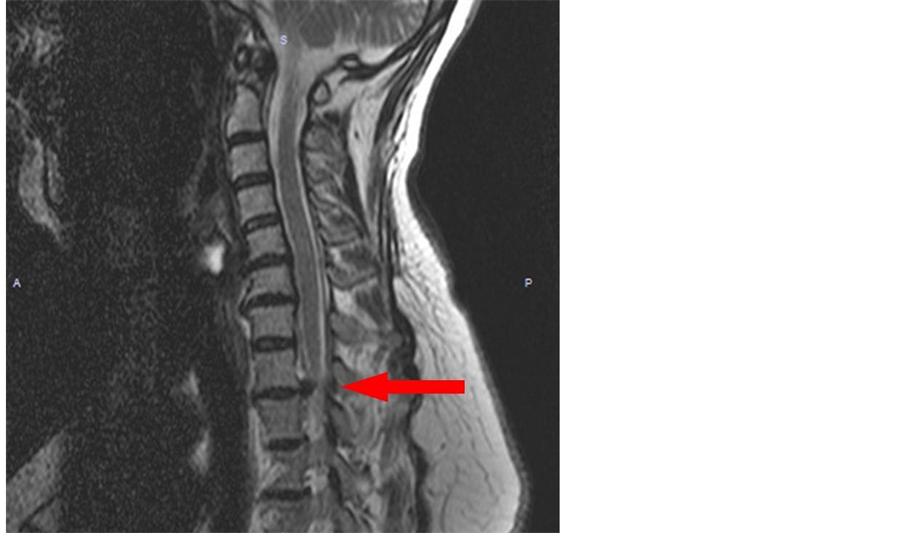
Figure 2. The arrow indicates the foraminal disc at T1-2.
Electromyography (EMG) showed denervation and neurologic motor unit potential changes, and minimal denervation of the inferior cervical muscles suggested a T1 radiculopathy.
Under general anesthesia, the patient was placed in the supine position. A transvers skin incision was made 2 cm above the right clavicle. After the discectomy, a right T1 foraminotomy was performed and a peak cage was inserted to the disc space. The postoperative period was uneventful. The pain radiating to the medial forearm and fourth and fifth fingers disappeared. The weakness of the hand improved to grade 4 within 2 months. The patient has no pain and regained muscle function in one year follow up.
3. Discussion
In the spine, T1-2 disc herniation is rare; similar compression at this level occurs with degenerative processes or adjacent segment disease following cervical fusion at neighboring levels [4] . Computed tomography (CT) and especially MRI can be used for the diagnosis [5] . The radiological examination must be performed carefully, because this area is generally at the caudal end of the range of cervical CT and MRI, and disc herniation may be easily missed [3] . T1 radiculopathy might accompany numbness of the fourth and fifth fingers or weakness of the intrinsic muscle of hand, similar to C8 radiculopathy. It can also produce Horner’s syndrome and sensory loss in the axilla, which are not found with C8 radiculopathy [1] [5] . Moreover, it can be confused with ulnar neuropathy, which is distinguished with EMG. In our case, EMG did not indicate an ulnar neuropathy.
Generally, T1-2 herniation causes T1 radiculopathy and, less often, myelopathy [1] [5] [6] . The outcome of surgery for T1 radiculopathy is mostly favorable [1] . Generally, T1-2 herniation is treated via a posterior approach, although this has some disadvantages. For example, excessive resection of the facet joint can cause instability resulting from hypermobility; there is limited surgical working space, and there is no chance of fusion in the disc space [6] . Rositti first described the anterior approach in 1993 [7] . Currently, most spinal surgeons prefer an anterior discectomy and fusion [8] . The anterior approach can be used to perform a simple discectomy and fusion, transcorporeally or via a microforaminotomy [6] -[9] . Preoperatively, the relationship between the sternum and T1-2 level must be investigated and the surgeon should be prepared to perform a sternotomy if required [3] . In our case, a sternotomy was not performed. In our opinion, the anterior approach is an easy, appropriate method for treating central and foraminal disc herniations. In addition, no sternotomy is required in many cases, especially for T1-2 disc herniation.
4. Conclusion
Careful clinical and radiological examination is very important for diagnosis. The anterior approach is an easy and appropriate method to treat central and foraminal disc herniation of the upper thoracic spine.
NOTES
*Corresponding author.