Microstructural Investigation of PM-355 Nuclear Track Detector Subjected to Low-Dose Gamma Irradiation: A Positron Annihilation Lifetime Study ()
1. Introduction
Polymeric materials are, at the present, extensively recognized as being of substantial meaning in a broad diversity of immense industrial and scientific interest [1]. Since radiation is one of the major factors that change the structural properties of polymers, in particular solid-state nuclear track detectors (SSNTDs), it would be meaningful to study the modification on their properties due to irradiation. Solid-state nuclear track detectors have found a variety of applications in different fields of science and technology [2]. These detectors have recently found extensive applications in plentiful high-temperature plasma experiments for corpuscular diagnostics. Diverse techniques have been applied in such studies and it has been widely established that the polymer CR-39 is a remarkably valuable radiation detector due to its capability to map out heavy ionized particles and neutrons [3-6]. An allyldiglycol polycarbonate (CR-39) plastic is one of the applicants for dosimeter material, having a dose response in an elevated gamma dose region. The particular CR-39 polymer PM-355 is of explicit concern, finding assorted applications in physical and technological sciences [7]. All versions of the detectors i.e. CR-39, PM-355 and PM-500 have identical chemical composition: C12H18O7 (polycarbonate of allyldiglycol). Differences among them are principally due to technical dealings such as additives of plasticizers, curing cycles and extrusion, quenching processes [8]. The PM-355 plastic appeared to be the most excellent one, particularly for the recognition of light ions including protons, deuterons, He-, C-, and S-ions, and consequently, this detector was principally used in the plasma experiments [9] and was suggested as an appropriate analytical tool for the laser-plasma experiment [10].
Irradiation by ionizing radiations is a distinctive and influential mean for modifying the physical properties of polymers. Radiation induces modifications in the chemical, physical and mechanical properties in the polymer material. Molecular chain scission, intermolecular crosslinking, rearrangement of bonding, and formation of carbon-rich clusters are some structural deformations in the irradiated polymer that direct to modify in the optical and dielectric properties of the material [11,12]. It is noticeable that the main physical interaction of radiations with the SSNTDs creates particular damage identified as chemical bond scission, free radicals and consecutive cross-linking [13]. For numerous polymers, together processes coexist and whichever one may dominate depending not only upon the chemical structure of the polymer, but also upon the circumstances of irradiation [14]. The cross-linking and main chain scissions that take place throughout irradiation possibly will lead to sharp changes in the physical properties of the polymer.
The microstructure of the sub-nanometer local freevolume holes that arise from the irregular molecular packing in polymers is largely determining the macroscopic properties of those materials. Positron annihilation lifetime spectroscopy (PALS) provides direct information about the size and numbers of microscopic holes in the polymer [15,16]. Therefore, in recent years, it has been used to explore the changes in the properties of irradiated polymer matrices. Positrons are injected in solid reach thermal energies in few picoseconds, and then after a diffusion path, they are trapped in open structures where they annihilate with an electron. A considerable fraction of the inserted positrons into the molecular materials, such as polymers, forms a positronium (Ps) [17] bound state. The annihilation characteristics are determined by the local electronic environment of the annihilation site. The bound state subsists both in triplet (orthopositronium, o-Ps) and in singlet (para-positronium, p-Ps) states, depending upon the spins of electron and positron, parallel or antiparallel, respectively. The comparatively long-lived ortho-positronium (o-Ps) experiences frequent collisions with molecules through which the positron of the Ps could annihilate with an electron other than its bound partner and opposite spin (pick-off annihilation). In amorphous part of polymers the o-Ps is preferentially formed and annihilated in the region of low electron density or in the so-called free volume holes [18,19]. This results in a exceedingly sensitive correspondence of the o-Ps pick-off rate and consequently the lifetime to the hole size [20,21]. The high specific trapping rate of positrons for open volumes makes positron annihilation technique a sensitive, non-destructive method for detection and characterization of open volume structures from single vacancies up to voids and porosities.
The studies of the irradiation changes taking place in the microscopic structure of materials are significant to material science. The aim of the present study is to investigate the changes induced by gamma irradiation doses from 1 × 10³ up to 9 × l03 Gray on the thermalproperties and microscopic structure of PM-355 SSNTDs. The study of the free volume presented in the amorphous phase was carried out by positron annihilation lifetime spectroscopy (PALS). The different techniques in the present work permitted us to get a complete depiction of the low dose gamma irradiation effects on the investigated SSNTDs.
2. Experimental
The PM-355 detectors used in the present study were procured by Rohm GmbH, USA and they are declared as “super-grade” AllylDiglicol Carbonate (CR-39). Samples having the dimension (1 × 1) cm2 and 0.3 mm thickness with density »0.90 gm/cm3 were prepared from a sheet of the super grade PM-355 detector. The chemical formula of all versions of the detectors i.e. CR-39, PM-500 and PM-355 has the same chemical composition: C12H18O7 (polyallyl-diglycol-carbonate). This formula was assumed according to the producer catalog [22]. The monomer contains two of the following functional groups; CH2 = CH-CH2. Differences between the detectors are principally due to technological procedures such as additives of plasticisers, curing cycles and extrusion, quenching processes [23]. The PM-355 samples were irradiated in the presence of air using a gamma-emitting source of 60Co. Doses between 1 and 9 kGy were applied at a fixed dose rate. The irradiation facility is constructed by the National Center for Radiation Research and Technology, Atomic Energy Authority of Egypt.
2.1. PAL
Positron lifetime measurements were then performed in these SSNTD at room temperature (about 25˚C) as a function of the gamma doses using a standard lifetime spectrometer, details of which are described elsewhere [24]. The principle of operation for the positron annihilation lifetime (PAL) spectrometer is to allow γ-γ coincidence measurements, positron emission should be accompanied by an appropriate start signal from a nuclear de-excitation like the 1.28 MeV photon in the case of the 22Na isotope. The 511 keV annihilation photon provides the stop signal. The PAL spectrometer consists of conventional fast-fast coincidence system with start and stop detectors, each of them made by coupling a fast (plastic) scintillator to a photomultiplier. The timing pulses are obtained by differential constant-fraction discrimination. The time delays between the start and stop signals are converted into amplitude pulses the heights of which are stored into a multichannel analyzer. Using 22Na, which is commercially available as an aqueous solution of 22NaCl, as a positron source. The source was prepared by evaporating a few drops of aqueous solution, about 15 μCi, of 22NaCl on a thin kapton foil of 12 μm thicknesses and an area of 10 × 10 mm2. After drying, 22NaCl spots were covered with another similar foil and properly sealed together with epoxy glue to avoid any leakage. Positrons emitted from the source had sufficient energy (Emax = 544 keV) to penetrate a certain depth of the sample. The source absorption by the kapton foil was about 7% and contributed to the medium lifetime components. This absorption was separated in the analysis of the lifetime spectra. The time resolution (full-width at half-maximum, FWHM) of the lifetime spectrometer was around 260 picoseconds (ps), as determined by the RESOLUTION computer code [25]. Each positron lifetime spectrum contains 106 counts which is sufficient to be analyzed with the computer program LT 9.0 in its distribution mode. The program gave three positron lifetimes as the best variance of fit.From a semiempirical model [26], o-Ps is assumed to be localized in an infinite high potential well with the radius rh + dr where rh is the hole radius and dr = 1.66 A˚ [15,20] describes the penetration of the Ps wave function into the hole walls. In this model the (mean) o-Ps lifetime  is used to calculate the mean radius
  of the holes (assumed spherical) by means of the equation
 (1)
(1)
where lpo is the pick-off annihilation rate of o-Ps inside the holes. The mean hole volume follows then from  Since the holes show a size and a shape distribution, a more reasonable way is to calculate the mean hole volume as mass centre of the hole volume distribution. LT9.0 assumes the annihilation rate distribution
  of the third annihilation channel to be log normal functions. Therefore the lifetime spectrum is expressed by the Laplace transformation of the functions
  where
where
 (2)
(2)
and

where  is the standard deviation of the distribution
 .
.
By determining the distribution  of the o-Ps annihilation rates and using Equation (1), the hole radius probability distribution
  can be calculated [27,28] as:
 (3)
(3)
where  is the free volume holes fraction with radii between
  and
and . The volume fraction of free volume holes with volume between
  and
and 
can be given by  while the fraction of holes with volume between
  and
and  could be given by
 .
.
2.2. TGA
The resulting o-Ps annihilation data will be correlated with the thermogravimetric analysis (TGA) of the PM- 355 samples. The analysis was carried out by TGA thermogram using a DuPont thermal analysis apparatus with temperature rate of 5˚C/min and temperature range from 20˚C to 500˚C.
2.3. Morphological Studies
The Conventional morphology observations of the PM- 355 samples were carried out by transmission electron microscopy (TEM) using JEOL JSM-1200 microscope operated at an acceleration voltage of 90 kV.
The outline of this research is exposing the PM-355 polymeric solid state nuclear track detector to low gamma radiation doses and investigate the changes induced by gamma irradiation on the microscopic structure of the PM-355 by using PAL, TGA, and TEM techniques.
3. Results and Discussion
3.1. Positron Annihilation Lifetime
All of the PAL spectra of the non-irradiated and irradiated PM-355 were analyzed assuming three discrete exponential components and the source correction due to annihilation in the source and source supporting Kapton [29]. Each lifetime corresponds to the average annihilation rate of a positron in a different state. The shortest lifetime component (τ1 = 0.12 - 0.15 ns, with intensity I1 = 24% - 43%) is related to the self-annihilation of p-Ps, atoms, while the intermediate one (τ2 = 0.364 - 0.417 ns and I2 = 39% - 56%) to annihilation of free positrons in the polymer matrix. τ2 is usually associated with the annihilation of positrons with valence or core electrons of the constituent atoms .The longest-lived component (τ3 = 2.071 - 2.328 ns and I3 = 12.2% - 29.2%) is associated with annihilation of o-Ps in the intermolecular spaces of polymer structure via the pick-off annihilation [30]. Taking τ3 as a measure of the free volume hole size and I3 as a number of free volume holes, the lifetime of o-Ps and its intensity for the non-irradiated sample have been deduced to be τ3 = 2.156 ± 0.026 ns and I3 = 29.2% ± 1.2%.
The positrons have a tendency to localize in comparatively more ordered regions, while Ps atoms tend to be localized in the disordered parts of a polymer matrix, and in semi-crystalline polymers Ps is localized in the amorphous regions and crystalline-amorphous interface [30].
Figure 1 shows the second lifetime τ2 and its relative intensity I2 of PM-355 samples as a function of gamma dose. The middle positron lifetime τ2 can be connected with positron annihilation on structurally intrinsic free volume traps, such as extended vacancy-like clusters [31]. The unirradiated sample had a τ2 of about 402 ps, so the vacancies in the sample are called vacancy clusters [32]. It is obvious from the figure that defect lifetime τ2 decreases after gamma irradiation. However, the relative intensity I2 increases with increasing irradiation dose. This implies that the concentration of vacancy clusters increased after irradiation [33]. During irradiation, both vacancies and interstitials are generated. The interstitials migrate to the vacancy clusters and diminish the size of vacancy clusters, so the defect lifetime τ2 decreases after gamma irradiation. More supersaturated vacancies are generated with increasing irradiation dose, the vacancies migrate and agglomerate, so the concentration of vacancy cluster decreased with increasing dose from 4 kGy up to 9 kGy.
The lifetime τ3 associated with the pick-off process as a function of gamma irradiation doses exhibits an increase up to 2.328 ns with an inflection point at the gamma dose 4 kGy as shown in Figure 2. The observed decrease in τ3 values with increasing dose may be attributed to a gradual formation of mobile charge carriers or

Figure 1. Variations of τ2 and I2 as a function of gamma radiation dose in PM-355 polymer.

Figure 2. Variation of the o-Ps lifetime, τ3, and its intensity, I3, with the irradiation dose for the PM-355 polymer.
easily orientable dipolar molecules. It is presumed that these charge carriers or dipolar molecules were formed from structural modifications (chain scission) caused by irradiation [34]. The free volumes are estimated using Equation (1) and found to decrease from 128.35 to 103.26 Å3.
Figure 2 shows the variations of the intensity of the o-Ps lifetime component I3 for gamma irradiation processes. The variations of I3 show a rapid decrease at the beginning up to 2 kGy. This decrease in I3 at low doses reflects a decrease in the number of free volume holes seems to fit quite well with the interpretation based on the process of cross-linking of the detector molecules. At higher gamma doses the scission of the molecular chains increases [35]. With increasing dose a smooth decrease is attained up to 7 kGy followed by leveling off up to 9 kGy, which is familiar for γ-irradiated polymers [36]. This behavior may indicate that stability in the structure has been achieved.
It is well known that when radiation impinging a polymer matrix, it leaves a trail of damage along its track in the form of broken molecular chains and free radicals. This behavior is quite reminiscent of what is expected by recombination and promoting important chemical and structural changes in the matrix. The inhibiting effect of radiation induced free radicals in polymers has been conjectured in some occasions. This is to elucidate the decrease in I3 with dose [37]. The cross-linking might take place due to the free radical recombination and the results indicated that the changes in the polymer properties depend on whether cross-linking or degradation dominates during the irradiation. An increase in crystallinity is effectively observed in the irradiation process where the decrease in I3 with dose is thus attributed to this effect [15]. The variation of I3 with dose established the trend observed for τ3 where the decrease in I3 could be persuaded by the change in the structural effect (i.e. an increase in the crystallinity degree with increasing dose).
The PM-355 has local molecular heterogeneity structure where the positron is annihilated therefore a continuous distribution of the inverse of the longest lifetime (τ3) can be obtained by analyzing the PAL spectra with the LT program. LT allows both discrete and log normal distributed annihilation rates λ = 1/τ. From the distribution of the o-Ps annihilation rate, λ3 = 1/τ3, the mean size and the size distribution of free volume holes can be calculated. Figure 3(a) shows the detected positron annihilation lifetime distribution for non-irradiated and irradiated samples. For non-irradiated sample, the free volume size is distributed from 78.65 to 146.19 Å3 (Full width at
 (a)
(a)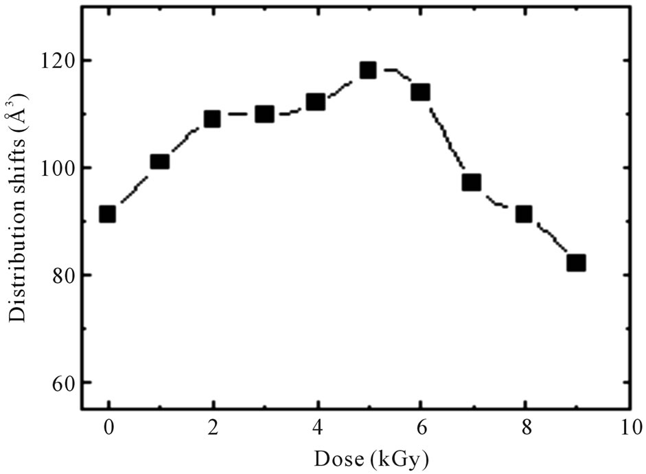 (b)
(b)
Figure 3. (a) Probability distribution function of unirradiated and irradiated PM-355 and (b) the shifts of the distribution function by gamma doses.
half maximum). Due to gamma-irradiation, the distribution shifts, Figure 3(b), to higher free volume size up to 5 kGy as a result of formation of new bonds or crosslinking which increase the o-Ps lifetime and after that the distribution shifts to lower free volume size up to 9 kGy. This is in agreement with the results obtained from the finite-term analysis.
3.2. Thermal Gravimetric Analysis (TGA)
To obtain a better understanding of the changes in thermal stability of PM-355 samples due to gamma irradiation TGA were performed in the temperature range from room temperature up to 500˚C, at a heating rate of 5˚C·min−1. The TGA thermograms for non-irradiated and irradiated samples are shown in Figure 4. The values of onset temperature of decomposition To were calculated by using these TGA thermograms, and are given in Figure 5 as a function of the gamma dose. The figure shows that To increases until a maximum value at 4 kGy irradiated sample indicating an increase in thermal stability of the polymer samples due to the cross-linking process, followed by a decrease with increase in gamma dose due to degradation (i.e. preferentially chain scission). The interpretation of these results may be that, in the dose range 1 - 4 kGy, the free radicals formed due to scission are chemically active and can be used in some chemical reactions that lead to the cross-linking mechanism. With increasing the gamma dose scission dominates which is reflected in a decrease in To of the PM-355 samples.
3.3. Transmission Electron Microscopy (TEM)
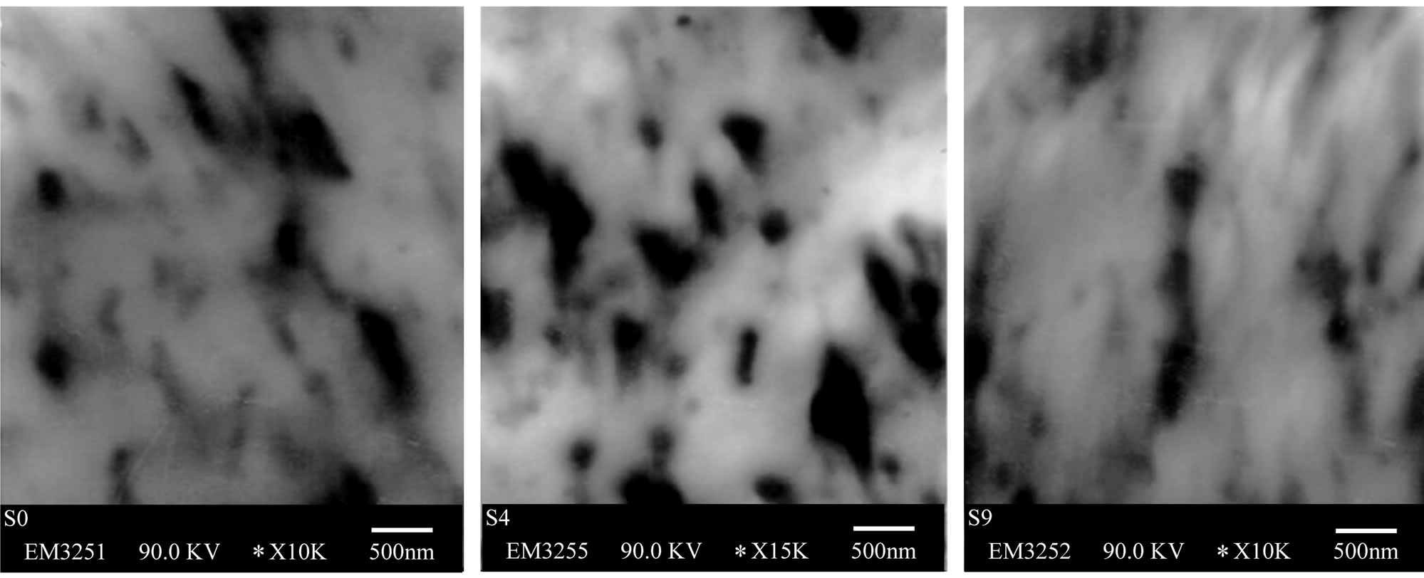
The micrographs of non-irradiated PM-355 SSNTD displayed structure of a semicrystalline polymer. The crystalline regions appeared as white areas as shown in Figure 6(a). The white areas are randomly oriented and

Figure 4. Thermogravimetric analysis of PM-355 as a function of gamma irradiation dose.
separated by highly entangled polymer molecules, dark regions, which form the amorphous phase. At low doses (4 kGy), gamma irradiation generates different free radicals which causes radiolytic scission of molecular chains by cleavage of C-C and C-H bonds. These free radicals experience recombination (leading to crosslinking) [38]. In the proposed microstructure for this stage, the crystallinity is less than in the virgin samples which could be deduced from the ×15,000 image as an increase in the dark regions concentration (see Figure 6(b)). Our TGA and PAL results on gamma-irradiated PM-355 confirm the diminution in crystallinity at 4 kGy. According to our measurements of TEM micrographs, the irradiated PM- 355 sample with 9 kGy dose (Figure 6(c)) showed an increase in crystallite region thickness in comparison with the 4 kGy sample. Thus, the increase in the crystallinity produced by the irradiation dose of 9 kGy was confirmed by the decrease in the decomposition temperature To and the ortho-positronium lifetime to-Ps.
4. Conclusion
From the results we have reported and discussed for the gamma irradiation on TM-355 polymeric detector, our conclusions can be summarized as follows. The defect lifetime τ2 decreases after gamma irradiation due to interstitials migration to the vacancy clusters and diminishes the size of vacancy clusters. With increasing irradiation dose, from 4 kGy to 9 kGy, the vacancies migrate and agglomerate, so the concentration of vacancy cluster decreases, which is confirmed by the I2 curve. The decrease of the free volumes from 128.35 to 103.26 Å3 is attributed to a gradual formation of orientable dipolar molecules due to structural modifications (chain scission)
caused by irradiation. The TGA thermograms show that the values of onset temperature of decomposition To increase until a maximum value at 4 kGy indicating an increase in thermal stability of the polymer samples. With increasing the gamma dose, scission dominates what is reflected in a decrease in To of the PM-355 samples. The TEM micrographs showed an increase in crystallite region thickness with 9 kGy dose sample in comparison with the 4 kGy sample. This increase in the crystallinity was confirmed by the decrease in the decomposition temperature To and the ortho-positronium lifetime to-Ps.
5. Acknowledgements
The author is thankful to Prof. Dr. S. A. Nouh, Ain Shams University, Faculty of Science, Physics Department
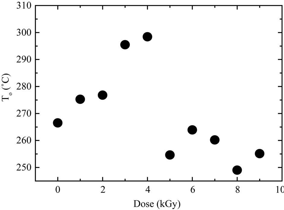
Figure 5. Effects of gamma irradiation on the decomposition temperature of PM-355.
 (a) (b) (c)
Figure 6. TEM image for (a) virgin PM-355, (b) 4 kGy gamma irradiated PM-355 and (c) 9 kGy gamma-irradiated PM-355.
for providing the samples. Thanks are also extended to the National Center for Radiation Research and Technology, Atomic Energy Authority of Egypt for providing the irradiation facility.