Controlled Growth of CdS Nanocrystals: Core/Shell viz Matrix
410
and cationic surface dangling bonds [10]. Zou, et al. [11]
have studied the effectiveness of various inorganic cap-
ping agents having different band gaps on the surface
passivation of cadmium sulphide (CdS) nanoparticles.
They have reported that it is possible to block the nonra-
diative channels on the surface of these nanoparticles by
capping them with wider band gap inorganic materials
like Cd(OH)2 and zinc sulphide (ZnS). It is also reported
that ZnS is more effective than Cd(OH)2 in surface pas-
sivation because of its better charge and size compatibil-
ity with CdS, resulting in increased band edge emission.
Growth of a wide band gap semiconductor ZnS on the
surface of a narrower band gap semiconductor CdS,
forming CdS/ZnS coreshell nanoparticles, leads to ap-
preciable passivation resulting in enhancement of pho-
toluminescence (PL) emission [14].
We reported synthesis of Mn2+ doped PVP-CdS nano-
matrix and CdS/ZnS core/shell nanostructures through
wet chemical approach. We capped CdS with both or-
ganic and inorganic capping agents. As organic is re-
sponsible for stroke’s shift where as inorganic gives bet-
ter enhanced luminescence efficiencies so we can choose
capping as per our requirement. After shell formation by
ZnS, monodisperse CdS nanoparticles were prepared,
which exhibited significantly enhanced luminescence
and high chemical stability. PVP role as a capping agent
is already discussed in our earlier paper [15].
2. Experiment Procedures
2.1. Sample Preparation
All the reactants and solvents were analytical grade. Cad-
mium acetate dihydrate Cd(CH3COO)2·2H2O (98%),
manganese acetate Mn(CH3COO)2 (98%), sodium sul-
fide (Na2S), zinc acetate Zn(CH3COO)2 (99.99%), were
purchased from Aldrich and used to synthesize or passi-
vate the CdS:Mn nanocrystals. Each (Cd2+, Zn2+, Mn2+)
and S2– containing standard aqueous solution was pre-
pared by dissolving Cd(CH3COO)2·2H2O, Zn(CH3COOH)2,
Mn(CH3CO O)2, and Na2S in water. The concentrations
of Cd2, Zn2+ and S2- in water were 0.1 M and the ratio of
Mn2+ to Cd2+ was fixed to 2 mol%.
Here we are going to report two synthesis processes.
In a typical preparation process 1.5 ml of 0.1M
Mn(CH3CO O)2 and 0.05 g PVP was dissolved in 25 ml
of 0.1M Cd(CH3COO)2·2H2O aqueous solution followed
by 15 ml of 0.1M sodium sulphide with continuous stir-
ring until a yellow solution of PVP capped CdS nanoma-
trix were formed. Where as in other set 2) 1.5 ml of 0.1M
Mn(CH3COO) 2 and 0.05 g PVP was dissolved in 25 ml
of 0.1M Cd(CH3COO)2·2H2O aqueous solution, then
15 ml of 0.1M sodium sulphide is added followed by
addition of 25 ml of 0.1M Zn(CH3COOH)2 with con-
tinuous stirring for favourable synthesis of Core/Shell
nanostructurs. Sequential addition of S2− and Zn2+ ions to
the (CdS)Mn core solution formed core/shell structures.
The obtained yellow solution was stored at roomtempe-
rature for measurements of optical absorption and pho-
toluminescence properties. Core/shell is a result of reac-
tion between Zn(CH3COOH)2 and excess Na2S, which
will lead to deposition of ZnS as a shell on the formed
core CdS nanoparticle.
2.2. Influence of Organic Capping
Two coordinating groups, nitrogen and carboxyl are pre-
sent in Poly Vinyl Pyrrolidone (PVP). Oxygen present in
PVP molecule makes coordinate bond with the Cd ions
whereas the lone pair of electrons on nitrogen in pyr-
rolidone is conjugated with the adjacent carbonyl group
and remaining oxygen in carboxylate makes coordinate
bond with the Mn ions. In PVP-CdS samples, we expect
a similar bonding at the nanoparticles, where in C=O-Cd+2
and C=O-Mn2+ bonds which can give rise to overlapping
of molecular orbitals of PVP with atomic orbitals of metal
ions. PVP as a capping agent plays a significant role not
only to increase the stability but also for the effective
doping of Mn into the CdS nanophosphors, which can be
attributed to the formation of coordinate bonding groups
between lone pair of oxygen atoms and those of metallic
atoms Cd and Mn in the CdS nanocomposite [15].
2.3. Characteristics Measurements
Transmission electron microscopic (TEM) photographs
were taken on Technai 30 G2 S-Twin. Ultraviolet–visi-
ble (UV-Vis) measured on Perkin–Elmer Mc Pherson
2035, Small angle scattering pattern were obtained by a
Rigaku D/max-2200 PC diffractometer operated at 40
kV/20 mA and Photoluminescence spectrophotometer
were measured on a Perkin–Elmer LS 55 spectropho-
tometer.
3. Result and Discussion
3.1. TEM Analysis
Sample preparation for TEM-Samples in mg were dis-
solved in ethanol and sonicated for 2 hrs then one drop of
this sample were taken on carbon coated copper grid, and
it was dried in oven for 24 hrs at 50˚C. Magnification is
200 KeV.
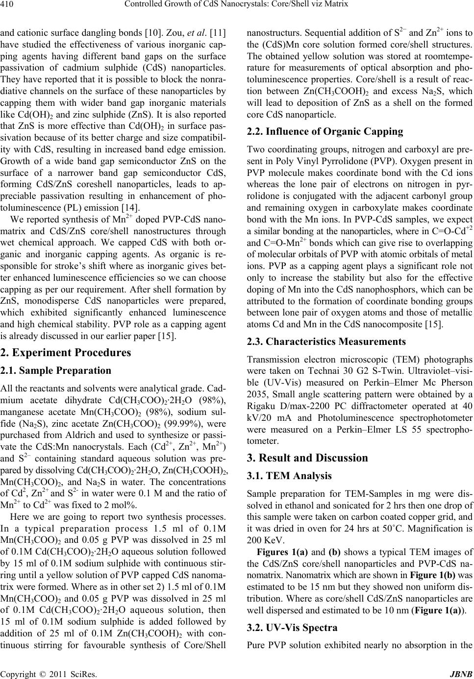
Figures 1(a) and (b) shows a typical TEM images of
the CdS/ZnS core/shell nanoparticles and PVP-CdS na-
nomatrix. Nanomatrix which are shown in Figure 1(b) was
estimated to be 15 nm but they showed non uniform dis-
tribution. Where as core/shell CdS/ZnS nanoparticles are
well dispersed and estimated to be 10 nm (Figure 1(a)).
3.2. UV-Vis Spectra
Pure PVP solution exhibited nearly no absorption in the
C
opyright © 2011 SciRes. JBNB


