Surgical Science, 2011, 2, 290-293
doi:10.4236/ss.2011.25062 Published Online July 2011 (http://www.SciRP.org/journal/ss)
Copyright © 2011 SciRes. SS
Endovascular embolisation of Giant Ruptured Proximal
Splenic Arterial Pseudoaneurysm Using Microcoils &
Glue: Case Report
Jawahar Rathod1, Kishor Taori1, Sachin Dhomane2, Prakash Pawar3, Devkaran Vaghasiya1,
Amit Disawal1, Amrita Guha1, Kiran Naiknavare1, Rahul Agale1, Prajwleet Gour1
1Department of Radio -diagnosi s , Government Medical College and Hospital, Nagpur, India
2Care Hospital, Nagpur, Indi a
3Sion Medical College & Hospital, Mumbai, India.
E-mail: drjawahar_12@rediffmail.com
Received January 10, 2011; revised April 15, 2011; accepted April 22, 2011
Abstract
Splenic artery aneurysms (SAA) are third most common intra-abdominal aneurysm (after aneurysms of the
abdominal aorta and iliac arteries). A splenic artery aneurysm larger than 3 cm is even rare. Splenic artery
aneurysms are fatal vascular lesions if ruptured. The incidence of rupture is increased in larger aneurysms.
Transcatheter arterial embolization is an alternative method of treatment in a patient with splenic arterial an-
eurysm which has relative lower morbidity and mortality than surgical procedures. Here, we report a case of
giant ruptured proximal splenic artery pseudoaneurysm detected by contrast enhanced computed tomography
(CECT) & referred to Interventional Radiology for Transcatheter arterial embolisation.
Keywords: Ruptured, Pseudoaneurysm, Microcoils, Glue, Pancreatitis
1. Introduction
Aneurysms of visceral arteries are found in less than 1%
of the general population [1 -4]. Splenic artery aneurysms
are the most common visceral artery aneurysms, ac-
counting for 60% to 71% [5-7], with a reported preva-
lence of 0.8% at arteriography [8]. SAA are more com-
mon in women than men. Pregnancy is the major risk
factor for rupture. Here, we report a case of acute on
chronic pancreatitits with g iant ru p tured proximal splen ic
artery pseudoaneurysm which was successfully em-
bolised using microcoils & cyanoacrylate glue.
2. Case Report
A 40-year-old male patient who was previously diag-
nosed as a case of acute hemorrhagic pancreatitits &
managed conserv atively 6 months back at our institu tion,
now came with h/o pain in epigastric region, hemateme-
sis & malena since 2 days. Laboratory tests revealed
slightly raised Sr. lip ase (214 U/L, normal: up to 190 U/L)
& anemia (Hb: 10.5 gm%). USG abdomen & Computed
tomography (CT) was performed, it showed changes of
acute on chronic pancreatitits (Figure 1) & a large spl-
enic artery pseudoaneurysm in its proximal part measur-
ing 5 × 3.8 cm. The aneurysm consists of peripheral
thrombosed part (Figure 2).
The patient was then referred to Interventional Radi-
ology department for emergency angio-embolisation
after giving Pneumococcal (pneumovac) and H. Influen-
zae B vaccination. Using Seldinger's method, via right
transfemoral approach, Selective angiography of the
splenic artery was performed using 5 Fr SIM 1 catheter
(cordis), It showed a large sessile pseudoaneurysm aris-
ing from the proximal segment of the splenic artery
(Figure 3). Through SIM 1 catheter, microcatheter
(Miraflex, 2.5 Fr) introduced into the splenic artery as a
co-axial system, tip of the microcatheter was placed be-
yond the pseudoaneurysm and embolisation started using
microcoils of various sizes (6 to 8 mm) but all the coils
(Total 8 coils) migrated at a single place of acute turn of
distal splenic artery. Angiography at this stage showed
patent aneurysm with embolisation of distal splenic ar-
tery. Then the decision was taken to embolise whole of
the splenic artery proximal to the microcoils using Glue
(N-butyl Cyanoacrylate). 6 ml 50% glue mixed with
J. RATHOD ET AL.291
Figure 1. NECT abdomen axial cuts reveals bulky hetero-
genous pancreas with multiple intra-pancreatic calcifications.
Figure 2. CECT abdomen axial cuts reveals giant saccular
anteriorly placed pseudoaneurysm (peripheral thrombosed
part & central patent part) arising from proximal splenic
artery.
lipiodol in equal proportion injected through the micro-
catheter upto the origin of the splenic artery. Bunch of
microcoils in distal splenic artery prevented glue from
entering into the splenic parenchyma. Post-embolization
angiogram & fluoroscopic image revealed successful
embolisation of the splenic artery with non-filling of the
pseudoaneurysm with microcils in the distal splenic ar-
tery & glue cast in the proximal splenic artery. (Figures
4 and 5)
3. Discussion
Splenic artery aneurysms are being diagnosed more com-
Figure 3. DSA angiogram of splenic artery showing pseu-
doaneurysm from proximal splenic artery
Figure 4. Post embolisation celiac angiogram showing pe-
ripheral bunch of coils with glue cast in the entire splenic
artery with patent hepatic & left gastric artery.
monly than in the past, mainly because of increased
availability of modern radiological imaging techniques
like computed tomography and angiography [5,6,9].
Treatment of splenic artery aneurysm is recommended in
patients with aneurysms greater than 2 cm [10]. Splenic
artery aneurysms are found most often in multiparous
women attributed to intimal hyperplasia. The prevalence
of splenic artery aneurysm has increased in patien ts with
portal hypertension and is estimated at 7% - 20% [11].
Copyright © 2011 SciRes. SS
J. RATHOD ET AL.
292
Figure 5. Post embolisation fluoroscopic image showing
peripheral bunch of coils with glue cast (arrow) in the
proximal splenic artery with patent hepatic & left gastric
artery.
Splenic artery pseudoaneurysm may be seen in patients
with trauma & pancreatitis. The splenic artery is the most
common site for pseudoaneurysms secondary to pan
creatitis [11]. Treatment of pseudoaneurysms is manda-
tory because risk of rupture is high [12,13]. These pa-
tients are often ill with severe pancreatitis and a mini-
mally invasive form of treatment would be of value.
Here we report a similar case in which the patient pre-
sented with splenic artery pseudoaneurysm which was
ruptured & presented with hematemesis & malena.. The
results of surgery for SAPAs are influenced by the pa-
tient's overall condition, and also by the anatomic loca-
tion of the pseudoaneurysm. Surgical mortality is ap-
proximately 16% for pseudoaneurysms in the head of the
pancreas and 50% for lesions in the tail [12]. Other un-
common causes of splenic artery aneurysm include fi-
bromuscular dysplasia, infection, and congenital anom-
aly [13]. Most aneurysms are small (2 cm in diameter),
saccular, and located at a bifurcation in a middle or distal
segment of the splenic artery [5,6,14]. The incidence of
rupture of splenic aneurysms is reported relatively less
frequently, occurring in 2% to 10% of cases, but the risk
increases when the aneurysmal diameter exceeds 3 cm,
when the size increases within a relatively short period,
or when the patient is pregnant [3,4,15]. Most splenic
artery aneurysms are detected incidentally during diag-
nostic imaging performed for other indications. Rupture
of splenic artery aneurysm is rare; however, the ruptured
splenic artery aneurysm left untreated is associated with
a high mortality rate [8]. In the present case report, the
size of the pseudoaneurysm was more than 3 cm & it was
ruptured. Treatment of splenic artery aneurysm is rec-
ommended in women of childbearing age, patients un-
dergoing liver transp lantation, patients with symptomatic
or expanding aneurysms, and patients with aneurysms
greater than 2 cm [8,10]. Endovascular methods are
coming to the forefront for the management of splenic
artery aneurysms. Splenic artery aneurysms away from
the hilum can be excluded with a stent graft. Emboliza-
tion of the entire splenic artery, if selective catheteriza-
tion of the aneurysm cannot be performed, is an alterna-
tive option [8,10]. We have also performed complete
embolisation of the splenic artery, distal & proximal to
the pseudoaneurysm using two different embolisation
materials, one is microcoils & another is liquid embolis-
ing material i.e. Glue for the first time. Surgery for
splenic artery aneurysms is associated with a mortality
rate of approximately 1%, but the mortality rate is in-
creased in patients with pancreatitis, in whom it is 16%
for those with aneurysms in the pancreatic head and 50%
for those with pancreatic body aneurysms. Splenic artery
aneurysms also may be treated with percutaneous inter-
ventional techniques such as transcatheter arterial em-
bolization, placement of a covered stent to exclude the
aneurysm, or percutaneous injection of coils or thrombin.
Transcatheter arterial embolization is associated with
significantly lower morbidity and mortality rate than
surgical procedures [16,17]. Embolization of intrasplenic
lesions may be performed with microcatheter-based
techniques, and success rates of 80% - 90% have been
reported for percutaneous transcatheter embolization[8].
Stent-graft placement across the aneurysmal neck to ex-
clude the aneurysm, a treatment that provides the benefit
of preserving blood flow through the splenic artery has
been reported [18,19]. In our case, we deployed the me-
tallic coils in the splenic artery distal to the aneurysm &
injected glue so that there was permanent complete oc-
clusion of the splenic artery & its aneurysm with no pas-
sage of cyanoacrylate glue into the distal most splenic
artery & its branches. It provides a cost effective method
to treat proximal splenic artery pseudoaneurysm. The
spleen has a rich vascular collateral supply, therefore
embolization is a more attractive treatment option th an it
would be in other visceral arteries. The occurrence of
complications after endovascular treatment is uncommon.
Possible complications include postembolization syn-
drome, transient elevation of pancreatic enzymes, splenic
infarction, infection, abscess, and, rarely, rupture of a
pseudoaneurysm8. Follow up imaging with CT or Dop-
pler ultrasonography is recommended for assessment of
the adequacy of treatment. The indications that warrant
surgical interventio n i.e. splenectomy after splenic artery
embolisation include hematologic complications and
Copyright © 2011 SciRes. SS
J. RATHOD ET AL.
Copyright © 2011 SciRes. SS
293
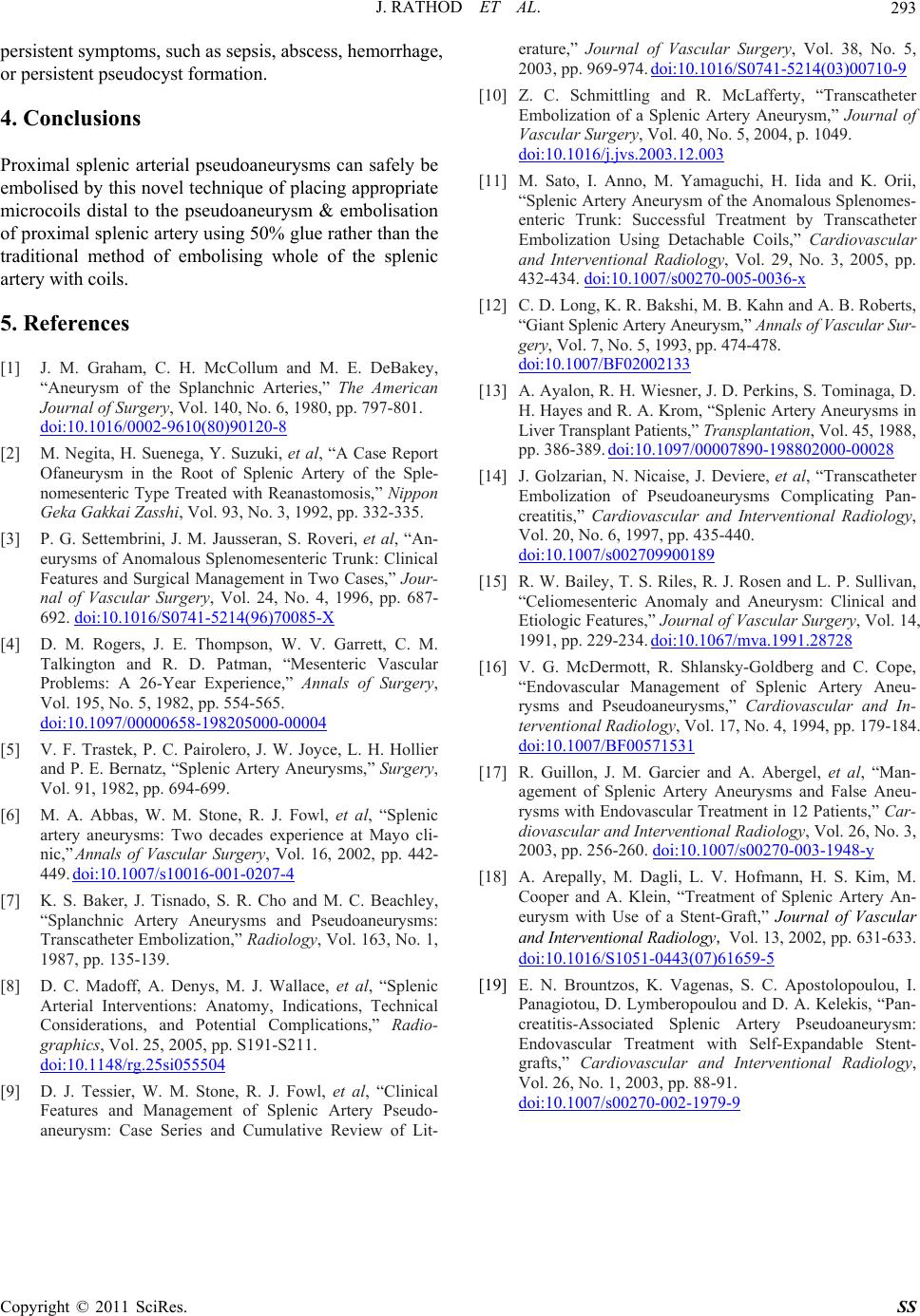
persistent symptoms, such as sepsis, abscess, h e morrhag e,
or persistent pseudocyst formation.
4. Conclusions
Proximal splenic arterial pseudoaneurysms can safely be
embolised by this novel technique of placing appropriate
microcoils distal to the pseudoaneurysm & embolisation
of proximal splenic artery using 50% glue rather than the
traditional method of embolising whole of the splenic
artery with coils.
5. References
[1] J. M. Graham, C. H. McCollum and M. E. DeBakey,
“Aneurysm of the Splanchnic Arteries,” The American
Journal of Surgery, Vol. 140, No. 6, 1980, pp. 797-801.
doi:10.1016/0002-9610(80)90120-8
[2] M. Negita, H. Suenega, Y. Suzuki, et al, “A Case Report
Ofaneurysm in the Root of Splenic Artery of the Sple-
nomesenteric Type Treated with Reanastomosis,” Nippon
Geka Gakkai Zasshi, Vol. 93, No. 3, 1992, pp. 332-335.
[3] P. G. Settembrini, J. M. Jausseran, S. Roveri, et al, “An-
eurysms of Anomalous Splenomesenteric Trunk: Clinical
Features and Surgical Management in Two Cases,” Jour-
nal of Vascular Surgery, Vol. 24, No. 4, 1996, pp. 687-
692. doi:10.1016/S0741-5214(96)70085-X
[4] D. M. Rogers, J. E. Thompson, W. V. Garrett, C. M.
Talkington and R. D. Patman, “Mesenteric Vascular
Problems: A 26-Year Experience,” Annals of Surgery,
Vol. 195, No. 5, 1982, pp. 554-565.
doi:10.1097/00000658-198205000-00004
[5] V. F. Trastek, P. C. Pairolero, J. W. Joyce, L. H. Hollier
and P. E. Bernatz, “Splenic Artery Aneurysms,” Surgery,
Vol. 91, 1982, pp. 694-699.
[6] M. A. Abbas, W. M. Stone, R. J. Fowl, et al, “Splenic
artery aneurysms: Two decades experience at Mayo cli-
nic,” Annals of Vascular Surgery, Vol. 16, 2002, pp. 442-
449. doi:10.1007/s10016-001-0207-4
[7] K. S. Baker, J. Tisnado, S. R. Cho and M. C. Beachley,
“Splanchnic Artery Aneurysms and Pseudoaneurysms:
Transcatheter Embolization,” Radiology, Vol. 163, No. 1,
1987, pp. 135-139.
[8] D. C. Madoff, A. Denys, M. J. Wallace, et al, “Splenic
Arterial Interventions: Anatomy, Indications, Technical
Considerations, and Potential Complications,” Radio-
graphics, Vol. 25, 2005, pp. S191-S211.
doi:10.1148/rg.25si055504
[9] D. J. Tessier, W. M. Stone, R. J. Fowl, et al, “Clinical
Features and Management of Splenic Artery Pseudo-
aneurysm: Case Series and Cumulative Review of Lit-
erature,” Journal of Vascular Surgery, Vol. 38, No. 5,
2003, pp. 969-974. doi:10.1016/S0741-5214(03)00710-9
[10] Z. C. Schmittling and R. McLafferty, “Transcatheter
Embolization of a Splenic Artery Aneurysm,” Journal of
Vascular Surgery, Vol. 40, No. 5, 2004, p. 1049.
doi:10.1016/j.jvs.2003.12.003
[11] M. Sato, I. Anno, M. Yamaguchi, H. Iida and K. Orii,
“Splenic Artery Aneurysm of the Anomalous Splenomes-
enteric Trunk: Successful Treatment by Transcatheter
Embolization Using Detachable Coils,” Cardiovascular
and Interventional Radiology, Vol. 29, No. 3, 2005, pp.
432-434. doi:10.1007/s00270-005-0036-x
[12] C. D. Long, K. R. Bakshi, M. B. Kahn and A. B. Roberts,
“Giant Splenic Artery Aneurysm,” Annals of Vascular Sur-
gery, Vol . 7, No. 5, 1993, pp. 474-478.
doi:10.1007/BF02002133
[13] A. Ayalon, R. H. Wiesner, J. D. Perkins, S. Tominaga, D.
H. Hayes and R. A. Krom, “Splenic Artery Ane urysms in
Liver Transplant Patients,” Transplantation, Vol. 45, 1988,
pp. 386-389. doi:10.1097/00007890-198802000-00028
[14] J. Golzarian, N. Nicaise, J. Deviere, et al, “Transcatheter
Embolization of Pseudoaneurysms Complicating Pan-
creatitis,” Cardiovascular and Interventional Radiology,
Vol. 20, No. 6, 1997, pp. 435-440.
doi:10.1007/s002709900189
[15] R. W. Bailey, T. S. Riles, R. J. Rosen and L. P. Sullivan,
“Celiomesenteric Anomaly and Aneurysm: Clinical and
Etiologic Features,” Journal of Vascular Surgery, Vol. 14,
1991, pp. 229-234. doi:10.1067/mva.1991.28728
[16] V. G. McDermott, R. Shlansky-Goldberg and C. Cope,
“Endovascular Management of Splenic Artery Aneu-
rysms and Pseudoaneurysms,” Cardiovascular and In-
terventional Ra diology, Vol. 17, No. 4, 1994, pp. 179-184.
doi:10.1007/BF00571531
[17] R. Guillon, J. M. Garcier and A. Abergel, et al, “Man-
agement of Splenic Artery Aneurysms and False Aneu-
rysms with Endovascular Treatment in 12 Patients,” Car-
diovascular and Interventional Radiology, Vol. 26, No. 3,
2003, pp. 256-260. doi:10.1007/s00270-003-1948-y
[18] A. Arepally, M. Dagli, L. V. Hofmann, H. S. Kim, M.
Cooper and A. Klein, “Treatment of Splenic Artery An-
eurysm with Use of a Stent-Graft,” Journal of Vascular
and Interventional Radiology , Vol. 13, 2002, pp. 631-633.
doi:10.1016/S1051-0443(07)61659-5
[19] E. N. Brountzos, K. Vagenas, S. C. Apostolopoulou, I.
Panagiotou, D. Lymberopoulou and D. A. Kelekis, “Pan-
creatitis-Associated Splenic Artery Pseudoaneurysm:
Endovascular Treatment with Self-Expandable Stent-
grafts,” Cardiovascular and Interventional Radiology,
Vol. 26, No. 1, 2003, pp. 88-91.
doi:10.1007/s00270-002-1979-9