Surgical Removal of Numerous Foreign Bodies from the Foot Caused by Sea Urchin Spines
98
Figure 2. Hand surgical instruments set. Instruments set
including: fine curved forceps, non-toothed forceps, skin-
hook, and a size 15 blade.
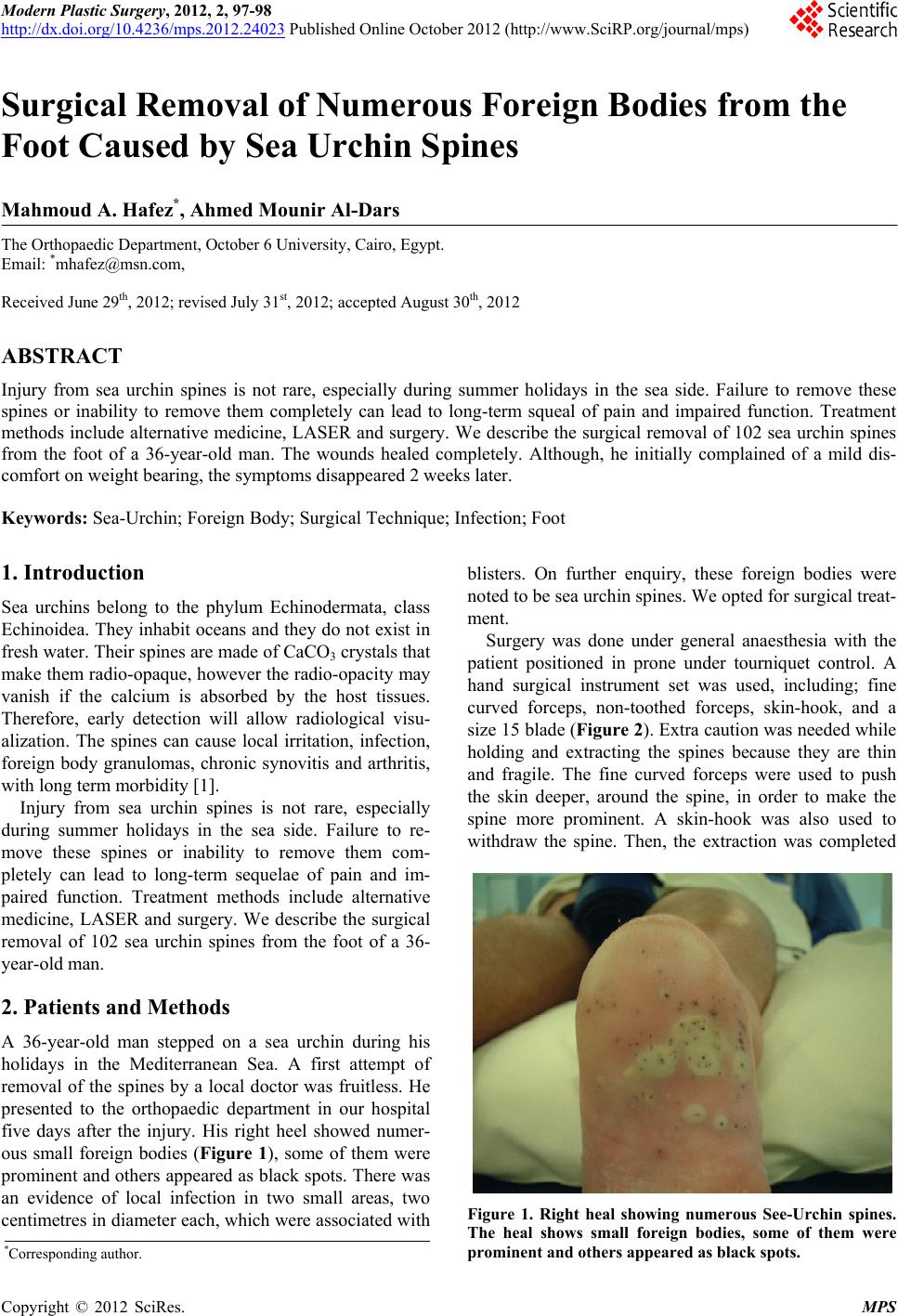
Figure 3. Removed foreign bodies. The foreign bodies were
counted as 102 including separated spines and buried
spines.
by gently using the either forceps. For the completely
buried spines, a small incision (0.5 cm) was made over
the black spot and then, a skin hook was used to with-
draw the spine as described above. The removal was
easier in the infected areas. It seemed that the blistering
resulted in partial withdrawal o f the spines from the deep
tissues. Complete excision of these blisters resulted in
easy removal of 60 spines. The time of surgery was 75
minutes.
3. Result
All foreign bodies were removed and we counted 102
spines (Figure 3). The patient had no postoperative
complications and he was discharged home the day after.
The wounds healed completely although he initially
complained of a mild discomfort on weight bearing,
hich disappeared at the last follow up, two weeks later.
The patient did not turn up in the later follow up appoint-
ments.
w
4. Discussion
Sea urchin spines can cause local irritation, infection,
foreign body granulomas. When they penetrate the der-
mis (or sub cutis). Newmeyer [2] reported that infection
could be due to the effect of their toxins, epithelial cov-
ering and any contamination from the water in which the
injury occurred. Removal of sea urchin spines is not
easy as the spines are small and they can easily break.
Several cases have been reported, describing different
techniques for removal. Falkenberg [3] reported a tech-
nique using alternative medicine by crushing the spines
in situ by stone then voiding fresh urine on the wound.
Burnett [4] reported the use of bolus ejection and Boer et
al. [5] reported the use of erbium: YAG LASER. New-
meyer [2] reported surgical removal of see urchin spines
from the hand.
The surgical technique we used is simple, aseptic, and
efficient and does not need sophisticated equipments.
The results were excellent and the spines could be re-
moved entirely even without radiological localisation.
Injury from sea urchin spines can cause pain and dis-
ability if left untreated. Awareness of the nature of this
injury and modalities of treatment is important to emer-
gency physicians and surgeons. Surgical removal is
recommended especially when there is infection. Su rgery
should be done under general anaesthesia, in theatre. The
surgeon should be prepared for a long and tedious task.
Fine instruments, good theatre light, tourniquet and sur-
gical loupe are invaluable.
REFERENCES
[1] W. J. Dahl, P. Jebson and D. S. Louis, “Sea Urchin Inju-
ries to the Hand: A Case Report and Review of the Lit-
erature,” Iowa Orthopaedic Journal, Vol. 30, 2010, pp.
153-156.
[2] W. L. Newmeyer 3rd., “Management of Sea Urchin Sp i ne s
in the Hand,” Journal of Hand Surgery—American Vol-
ume, Vol. 13, No. 3, 1988, pp. 455-457.
doi:10.1016/S0363-5023(88)80031-5
[3] P. Falkenberg, “Sea Urchin Spines as Foreign Bodies—
An Alternative Treatment,” Injury, Vol. 16, No. 6, 1985,
pp. 419-420. doi:10.1016/0020-1383(85)90062-2
[4] J. W. Burnett, “Bolus Ejection: A Method for Removing
sea Urchin Spines,” Annals of Emergency Medicine, Vol.
39, No. 1, 2002, pp. 94-95.
doi:10.1067/mem.2002.120747
[5] A. Boer, F. R. Ochsendorf, C. Beier and R. Kaufmann,
“Effective Removal of Sea-Urchin Spines by Erbium:
YAG Laser Ablation,” British Journal of Dermatology,
Vol. 145, No. 1, 2001, pp. 114-116.
doi:10.1046/j.1365-2133.2001.04306.x
Copyright © 2012 SciRes. MPS