Novel Technique Combining Tissue and Mesh Repair for Umbilical Hernia in Adults ()
1. Introduction
Repair of umbilical hernia in adults is one of the commonest procedures performed on middle aged population. The traditional Mayo’s repair is associated with increased failure rates. This has prompted surgeons to use mesh reinforcement. Laparoscopic approach has also emerged as one of the surgical options. However none of the aforementioned repairs can provide good long lasting results [1] . Therefore the need arises to develop a good repair which ensures a long term recurrence-free outcome.
1.1. Objective
Aim of the study was to develop a new technique which combines tissue repair with mesh reinforcement.
1.2. Inclusion Criteria
All adult patients diagnosed clinically with umbilical hernia.
■ 1.3. Exclusion Criteria
■ Patients who had undergone surgery previously around the umbilicus and had developed umbilical hernias.
■ Complicated umbilical hernias.
2. Material and Methods
The study was conducted in a single surgical unit of Dr. D. Y. Patil hospital and research centre in Navi Mumbai in the period from January 2012 to June 2012. The study protocol was discussed and prior approval of the hospital ethics committee was sought. Patients diagnosed as umbilical hernia with well controlled comorbidities if present were included in the study. Patients were admitted one day prior to surgery. On admission, a detailed written informed consent was sought from each patient included in the study prior to the surgery. Thereafter a detailed proforma was completed which included demographic data and clinical details. Perioperative antibiotics were administered comprising 3 doses of intravenous Ceftriaxone 1 gm and Amikacin 500 mg (i.e. pre op, intra op and post op). All procedures were carried out under general anaesthesia. Patients were discharged after removal of drains and followed up.
3. Surgical Technique
All cases were operated upon by the first author. (KV) A vertical incision extending one inch above umbilicus to an inch below umbilicus, curving along the umbilicus to one side was made. The umbilical skin was dissected free from the underlying sac. The sac was dissected till the neck which was identified by a thick fibrous ring. The sac was opened, contents reduced and herniotomy performed (Figure 1). Two incisions were made 1 cm from midline on either rectus sheaths and flaps were created. The vertical extent of the incisions was 1 inch above to 1 inch below the level of the defect. These flaps were approximated in the midline with a non-absorbable suture material (Figure 2). A segment of the rectus muscle underlying these flaps was dissected free from the posterior rectus sheath in order to create a retro-rectus space. Polypropylene mesh altered to the size of the defect with an extra inch all around the defect was placed over newly created midline. It was fixed in midline and laterally with interrupted non-absorbable suture (Figure 3).

Figure 1. The defect is outlined by the black circle. The blue arrows point to the edges of the defect.

Figure 2. Reconstituted midline marked by the purple line after approximation of the medical cut edges of the anterior rectus flaps marked by blue arrows. The black arrows point towards the lateral cut edge of the anterior rectus sheath with the underlying rectus abdominis muscles on either side.
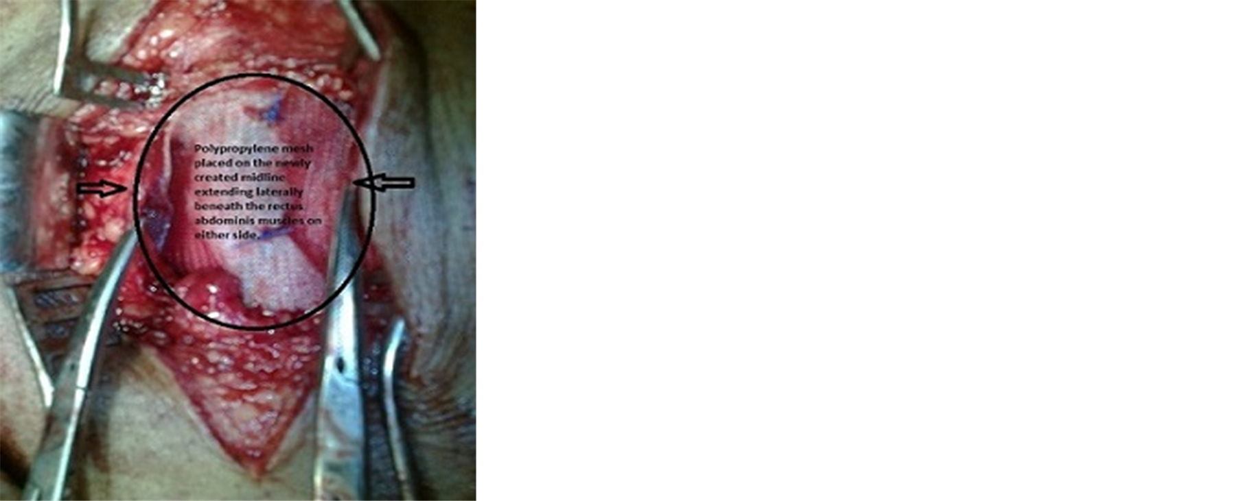
Figure 3. Mesh place over the newly created midline and extending underneath the rectus abdominis muscles on either side marked by the black arrows.
A negative suction drain was placed over mesh and brought out through a separate incision. The lateral cut edge of the anterior rectus sheath of either side was approximated with interrupted No. 1 Ethilon stitches (Figure 4). Subcutaneous tissues were approximated with absorbable suture taking utmost care to reconstitute the umbilical skin. Skin edges were approximated using a stapler.
Drains were removed after 48 hr and patient discharged thereafter. Skin staples were removed on 10th postoperative day. Patients were advised to use an abdominal binder for 12 weeks (Figure 5).
4. Results
The results of the study were tabulated (Table 1). The mean age of patients was 48.8 ± SD of 5.5 yrs. (range 38 - 57 years). There were 17 females and 3 males who underwent this procedure. The mean BMI was 26.5 (range 25 - 29). Comorbidities observed in these patients were diabetes mellitus (DM) in 4, hypertension (HTN) in 4, IHD in 4 and 1 patient had ascites. 3 patients had all 3 comorbidities (DM, HT, and IHD) and 2 out of these 3 patients developed superficial wound infection, whereas 2 patients had HT and IHD and 1 patient had DM

Figure 4. A negative suction drain placed over the mesh and brought out through a separate incision. The lateral cut edges of the anterior rectus sheath approximated with non absorbabale sutures marked by the black arrows.
with ascites. The mean stay of patients in hospital was 3.3 days (range 3 - 5 days). The mean follow up was 13 months (10 - 18 months). There was no recurrence in any of the patients with this technique.
5. Discussion
Attempts to evolve a standardized repair for umbilical hernia in the adult population still continue. A variety of techniques were developed for the repair [1] [2] . However most of these techniques did not sustain for long and gradually became obsolete. Obesity which has become a global epidemic especially in the urban population has become the biggest impediment to a successful outcome. Obesity happens to be important and significant factor in addition to age in development of umbilical hernias. Advancing age accompanied with obesity significantly predisposes to development of umbilical hernias. Umbilical hernias are also more commonly seen in female population. Weakening of abdominal wall after pregnancy heightens the incidence of umbilical hernia in women. This was observed in current study wherein the mean age of the patients was 48.8 yrs (± SD of 5.5). 85% of patients in the present study were women and majority were obese (High BMI). Comorbidities such as DM, HT, IHD, ascites may impact surgical outcomes. This may be due to poor wound healing in diabetics and increased chances of developing hematomas in hypertensive patients. Patients with IHD may have ascites as seen in cardiac failure accompanied with poor vascularity of local tissues. Therefore developing a new technique needs to take into consideration intrinsic tissue factors which led to the development of the hernia along with extrinsic factors which create impediments to successful outcomes.
The traditional Mayo’s repair comprises of horizontal double breasting of tissues withstood the test of time

Table 1 . Results of the case series.
(BMI: Body mass index; DM: Diabetes mellitus; HT: Hypertension; IHD: Ischaemic heart disease; Other: Ascites; F: Female; M: Male; +: Present; -: Absent).
for patients who are not obese. However in the obese subgroup of patients the failure rate with this technique started rising thereby prompting surgeons to devise another repair. The use of a mesh was therefore advocated. [2] [3] . The mesh however had its intrinsic complications. Because of anatomical intricacies of umbilical region, meshes were placed on the defect by onlay technique. This led to complications ranging from irritation caused by mesh to infections. Infections in the peri-umbilical region are common despite adequate prophylaxis. Infection developing in a hernia repair leads to complete failure with significant morbidity and cost implications [4] [5] .
The advent of laparoscopy led to development of a technique for repair of umbilical hernias. Special non-absorbable adhesion-free meshes were placed intraperitoneally and fixed with tacks [2] [6] [7] . This approach has significant draw backs. Dissection of the sac may at times be difficult prompting conversion to open. The fibrous defect remains unobliterated and is just covered by mesh from within. There is high incidence of loosening of tacks thereby leading to collapse of mesh within peritoneal cavity. This predisposes to significantly morbid adhesive intestinal obstruction [6] [7] .
Pain following laparoscopic umbilical hernia repair is a very morbid condition due to wide spread use of tacks. Laparoscopy does not offer any cosmetic advantage as the redundant umbilical skin remains untouched. Therefore laparoscopy as a procedure for umbilical hernia repair is not an attractive option as it lacks sound technical and cosmetic fundamentals [6] .
The procedure presented in the study is based on the assumptions that in hernia patients there is a weakening of both the local aponeurotic structures and the process of healing.
The umbilicus is a potential weakness in the anterior abdominal wall. Exposure to high intra-abdominal pressure predisposes to give way of the umbilical cicatrix. This happens usually in the midline just above or below umbilical cicatrix. Hence, these hernias are designated as paraumbilical hernias in adults. The hernia sac which usually forms has a narrow neck thereby predisposing to complications. Therefore it is always advisable to repair such hernias at the earliest. As there is deficient midline in these patients at the site of herniation, it is essential to construct a midline [8] . This midline is created from flaps of anterior rectus sheath [8] [9] . Approximation of these flaps provides a strong midline (Figure 2). However as the tissues are intrinsically weak one cannot rely solely on the new midline created by these flaps [10] [11] . Thus it is prudent to reinforce the newly created midline with a mesh [12] -[15] . Therefore polypropylene mesh tailored to the size of the defect in each patient is used and fixed both in the midline and laterally. Laterally the mesh lies below the rectus abdominis muscles on either side (Figure 3). The space containing the mesh and rectus muscles is closed by the approximation of the lateral cut edges of anterior rectus sheath. A negative suction drain is placed over the mesh within the closed space in order to prevent development of hematomas and seromas which could predispose to increase tension within the space leading to break down of sutures [15] (Figure 4). The mesh remains sandwiched between anterior and posterior rectus sheath thereby reducing the chances of infection significantly [15] .
The subcutaneous tissue needs to be approximated meticulously taking into consideration the cosmetic implications of the newly formed umbilicus [16] [17] . Skin is approximated with staples which help in providing a fine scar. This technique preserves the umbilicus which is a very important concern in female patients (Figure 5). 2 patients with significant comorbidities developed superficial wound infection which were cured by dressings only without any damage to the underlying repair. The mean hospital stay of patients was 3.35 days which is comparable to other studies [18] [19] . Patients were pain-free at the time of discharge. Mean follow up was 13 months with no recurrence in any of the patients. We therefore advocate this repair for umbilical hernias in adults as it has no recurrence rate. This repair is financially cheaper than a laparoscopic repair as the mesh used in this repair is an ordinary mesh unlike the one used during laparoscopic repair [20] . This is an added advantage for patients in the developing world where cost is a very important determinant of the approach to be used. The limitation in the study was the sample size. The technique needs to be carried out on a large sample size followed by a prospective randomized trial to weigh the benefits over other forms of repair.
6. Conclusion
This novel technique of mesh reinforcement of tissue repair is best suited for repair of umbilical hernias in adults in view of excellent results and low costs.
Acknowledgements
We would like to thank Dr. Shirish Patil, Dean of Dr. D. Y. Patil Medical College, Navi Mumbai, India for allowing us to publish this study. We would also like to thank Mr. Parth K. Vagholkar for his help in typesetting the manuscripts.
Funding: Nil.
Conflict of interest: Nil.
NOTES
*Corresponding author.