Reversed-Phase HPLC Analysis of Steviol Glycosides Isolated from Stevia rebaudiana Bertoni ()
1. Introduction
Recent interest of food and beverage industry has driven their focus towards natural high-potency sweeteners due to the increasing awareness of obesity problem and the health impacts associated with certain artificial sweeteners. Hence many soft drink manufacturers are trying to reduce calories by introducing natural non-caloric sweeteners into their systems. Stevia rebaudiana Bertoni, a perennial shrub belong to the family of Asteraceae (Compositae) is native to Paraguay and Brazil, but now is grown commercially in a number of countries, particularly in Japan, Taiwan, Korea, Thailand and Indonesia [1] [2] . Extracts of the leaves of S. rebaudiana have been used for decades to sweeten food and beverages in Japan, South America and China is one such example for the source of natural high potency sweeteners. The major constituents in the leaves of S. rebaudiana are the potently sweet glycosides namely stevioside, and rebaudiosides A; which are glycosides of the diterpene ent-13-hydroxykaur-16-en-19-oic acid and also known as steviol [3] [4] . Stevioside tastes about 150 - 250 times sweeter than sucrose, whereas rebaudioside A tastes about 200 - 300 times sweeter than sucrose. Both are noncaloric. These compounds are also known as stevia sweeteners.
As a part of our research related to the discovery of natural sweeteners and sweetener enhancers as well as developing analytical methods to identify and separate them, we are herewith describing a qualitative HPLC method for the nine steviol glycosides reported in Joint FAO/WHO Expert Committee on Food Additives (JECFA) 2010 monograph [5] namely rebaudioside A (1), steviolbioside (2), stevioside (3), rubusoside (4), rebaudioside B (5), rebaudioside C (6), rebaudioside C (7), rebaudioside D (8), and dulcoside A (9) using a condition closely associated with JECFA at temperatures 20˚C, 40˚C, 60˚C, and 79˚C. Using this method, the retention times of nine various steviol glycosides 1-9 (Figure 1) were identified at four temperatures 20˚C, 40˚C, 60˚C, and 79˚C. Also, calculated the relative retention times for the steviol glycosides 2-9 against the standard steviol glycoside, rebaudioside A (1). Recently, there are several reports on the isolation, spectral analysis and analytical methods reported in the literature for the steviol glycosides isolated from S. rebaudiana [6] -[12] .
2. Experimental
2.1. Reagents and Chemicals
Stevia glycoside standards kit containing dulcoside A, steviobioside, stevioside, rebaudioside A, rebaudioside B, rebaudioside C, rebaudioside D, rebaudioside F, rubososide was obtained from Chromadex (P/N KIT-00019568- 010, Irvine CA). HPLC grade acetonitrile and water were obtained from Fischer Scientific (Fair Lawn, NJ), and Pharm Co. (Brookfield, CT). Phosphoric acid (49% - 51%) HPLC grade was acquired from Sigma-Aldrich (Bellfonte, PA) whereas Sodium phosphate (monobasic) laboratory grade was obtained from Scientific Strategies (Oklahoma, OK).

Figure 1. Structures of ent-13-hydroxykaur-16-en-19-oic acid glycosides (1-9).
2.2. Mobile Phase Preparation
All solvents were degassed for at least fifteen minutes before use. The HPLC method employed was an isocratic binary solvent mobile phase system with a 32:68 mixture of acetonitrile and 10 mmol/L sodium phosphate buffer (pH: 2.63), very similar to the method reported in JECFA [5] .
2.3. Standard Preparation
Each standard steviol glycoside 1-9 was prepared separately at a concentration of 10 mg/ml. The dilutions for all steviol glycosides (1, 3-4, 6-9) were made using the phosphate buffer, but for steviolbiososide (2) and rebaudioside B (5) were prepared in methanol. Then the mixture of standard steviol glycosides 1-9 was prepared such that dilutions were made to set the concentration at 0.25 mg/L of each compound in the mixture.
2.4. Instrumentation and Conditions
An Agilent (Wilmington, DE) 1100 HPLC System, including a quaternary pump, a temperature controlled column compartment with additional 6 port switching valve, an auto sampler and VWD absorbance detector, was used for analysis. The detector was set-up at UV 210 nm. The data acquisition was done using a Chemastation A 10.02 software. The column used for HPLC analysis was a reversed-phase C18 (2) 100 A Phenomenex (Torrance CA) (Length: 250 mm, inner diameter 4.6 mm, particle size: 5 µm); pH was measured using meter Metler Toledo seven compact pH/ion S220 (Switzerland). Branson Ultrasonic Cleaner Model 2510 (Maplewood, NJ) was used for degassing HPLC solvents.
2.5. Analysis Procedure
For the RP-HPLC method, the column was flushed with 50 mL of 90% MeCN to waste before use and the samples were bracketed with standards by injecting them at the beginning and at the end of a run for accuracy of their retention times. 5 µL of steviol glycoside mixture (1-9) has been injected at temperatures 20˚C, 40˚C, 60˚C, and 79˚C. All the nine steviol glycosides were detected under UV at 210 nm. The two compounds rebaudioside A (1) and stevioside (3) were injected once at the beginning and once at the end of the sequence for consistency.
3. Results and Discussion
Generally separation of stevia sweeteners whose main difference from the chemical structural point of view is the number and type of glycoside moieties attached to aglycone moiety steviol, will be carried out using an amino (NH2) column. Using this method, major steviol glycosides present in stevia extracts or purified steviol glycosides can be identified easily. Since water is a strong solvent for NH2 columns, a reversed-phase C18 column is generally preferred due to their superior reproducibility and durability. However, on a standard C18 column, some of the steviol glycosides elute at same retention times (tR), and with recently developed stationary phases addressed the limitations of the older bonded polar and bonded non-polar phase columns. Detection sensitivity was also a key factor considered in the development of HPLC method. In order to develop a suitable method for the separation of the glycosides, we have tested various conditions and found that although these are weak UV absorbers, they give an adequate signal at 210 nm to meet the required quantitation limit (LOQ) of 0.5 mg/L.
In this study, we have utilized an HPLC method with a reversed-phase column under isocratic binary solvent mobile phase system (32:68 mixture of acetonitrile and phosphate buffer) at temperatures 20˚C, 40˚C, 60˚C, and 79˚C. The pH of the buffer used in the HPLC method was at 2.63 and the injection volume of the standard steviol glycoside mixture was 5 µL. Duplicate runs were performed at each temperature and found almost identical results; hence one representative HPLC chromatogram for the mixture of steviol glycoside 1-9 at four different temperatures 20˚C, 40˚C, 60˚C, and 79˚C is given in Figures 2-5.
The retention times for all the 9 naturally occurring steviol glycosides rebaudioside A (1), steviolbioside (2), stevioside (3), rubusoside (4), rebaudioside B (5), rebaudioside C (6), rebaudioside C (7), rebaudioside D (8), and dulcoside A (9) were identified and are given in Table1 A close observation of the retention times from the above Figures 2-5 and Table 1 indicated that better separation of steviol glycosides 1-9 was observed at higher temperatures.
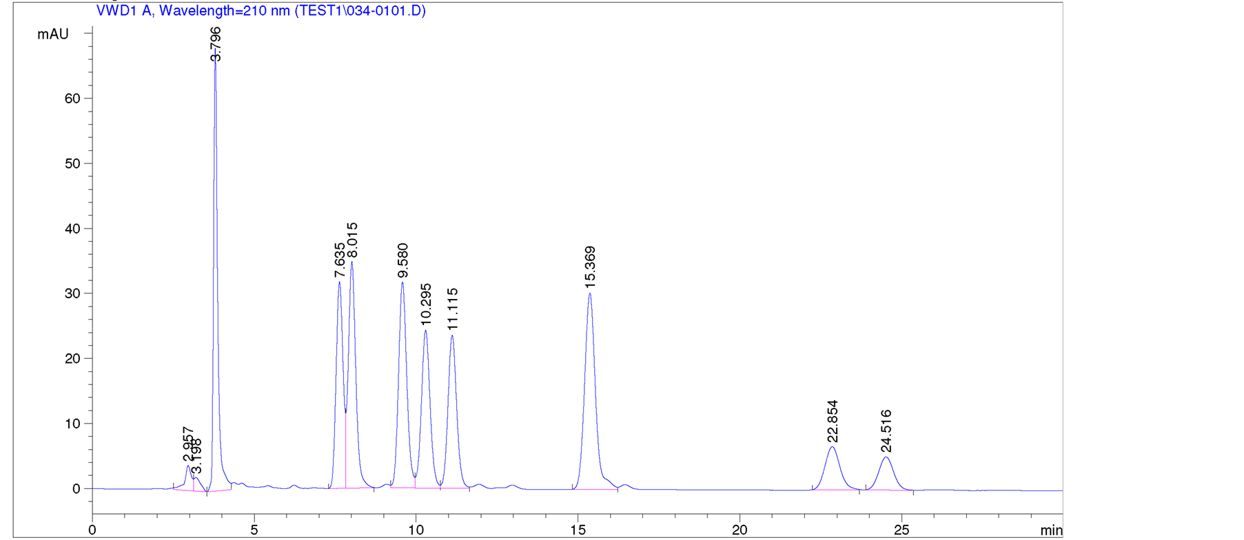
Figure 2. HPLC chromatogram for steviol glycosides 1-9 at 20˚C.

Figure 3. HPLC chromatogram for steviol glycosides 1-9 at 40˚C.
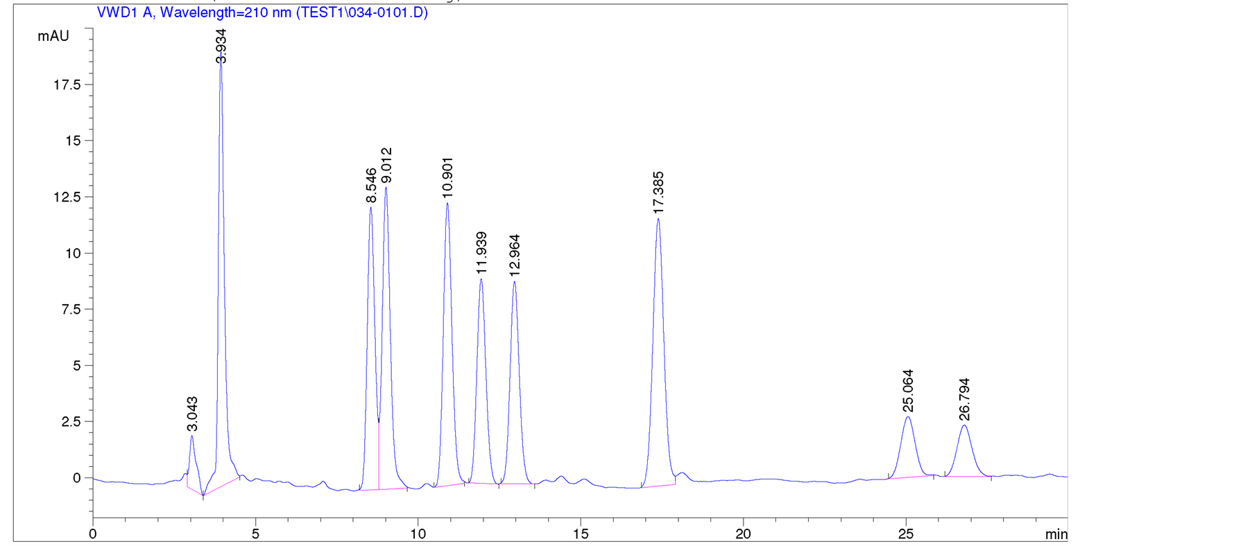
Figure 4. HPLC chromatogram for steviol glycosides 1-9 at 60˚C.
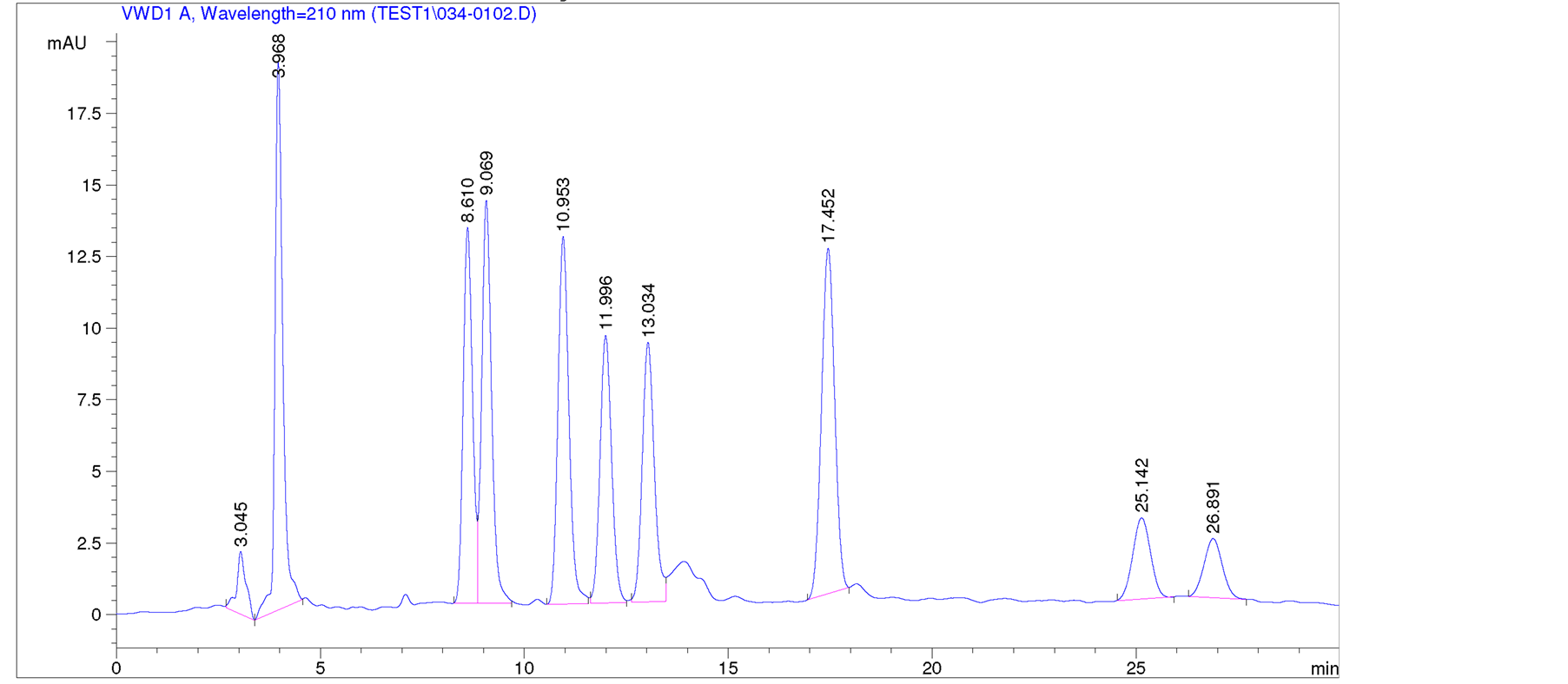
Figure 5. HPLC chromatogram for steviol glycosides 1-9 at 79˚C.

Table 1. Retention (tR) times of steviol glycosides 1-9 at temperatures 20˚C, 40˚C, 60˚C, and 79˚C.
The two major steviol glycosides of S. rebaudiana rebaudioside A (1) and stevioside (3) and the three minor compounds rebaudioside C (6), rebaudioside F (8), and dulcoside A (9) were having good separation at higher temperatures 40˚C, 60˚C, and 79˚C. The other four minor steviol glycosides steviolbioside (2), rubusoside (4), rebaudioside B (5), and rebaudioside C (7), which were having retention times not close to the other five steviol glycosides separated well at all temperatures. From the above experiments it has been concluded that temperatures 40˚C and 60˚C would be ideal for steviol glycoside separation, whereas some degradation appeared at 79˚C. Currently, several other experiments are in progress to find out the best conditions to separate various steviol glycosides for their isolation using HPLC by changing the flow rate, pH, as well as finding the best temperature between 40˚C and 60˚C.
Also, the relative retention times for the steviol glycosides 2-9 were calculated against the major steviol glycoside, rebaudioside A (1) and are given in Table2
4. Conclusion
We are herewith reporting a qualitative HPLC method for the identification of nine steviol glycosides—rebaudioside A (1), steviolbioside (2), stevioside (3), rubusoside (4), rebaudioside B (5), rebaudioside C (6), rebaudioside C (7), rebaudioside D (8), and dulcoside A (9) based on the RP-HPLC method using UV detection system. Experimental results suggested that temperatures 40˚C and 60˚C would be ideal for steviol glycoside separation, whereas it was found that the steviol glycosides did not resolve clearly at 20˚C, and some degradation at 79˚C.
Table 2. Relative retentions (RRT) of steviol glycosides 2-9 against rebaudioside A (1).
Acknowledgements
We thank James May, Chief Executive Officer and Carol May, President of Wisdom Natural Brands for their support.
NOTES
*Corresponding author.