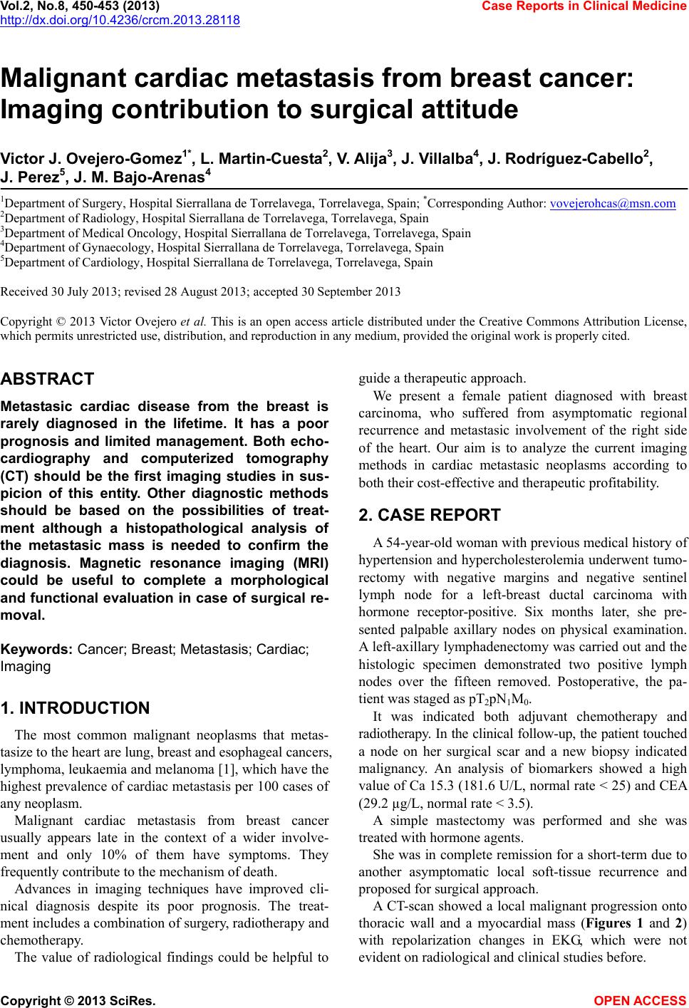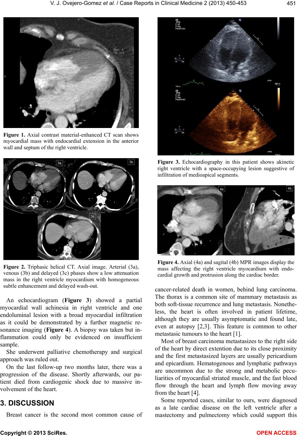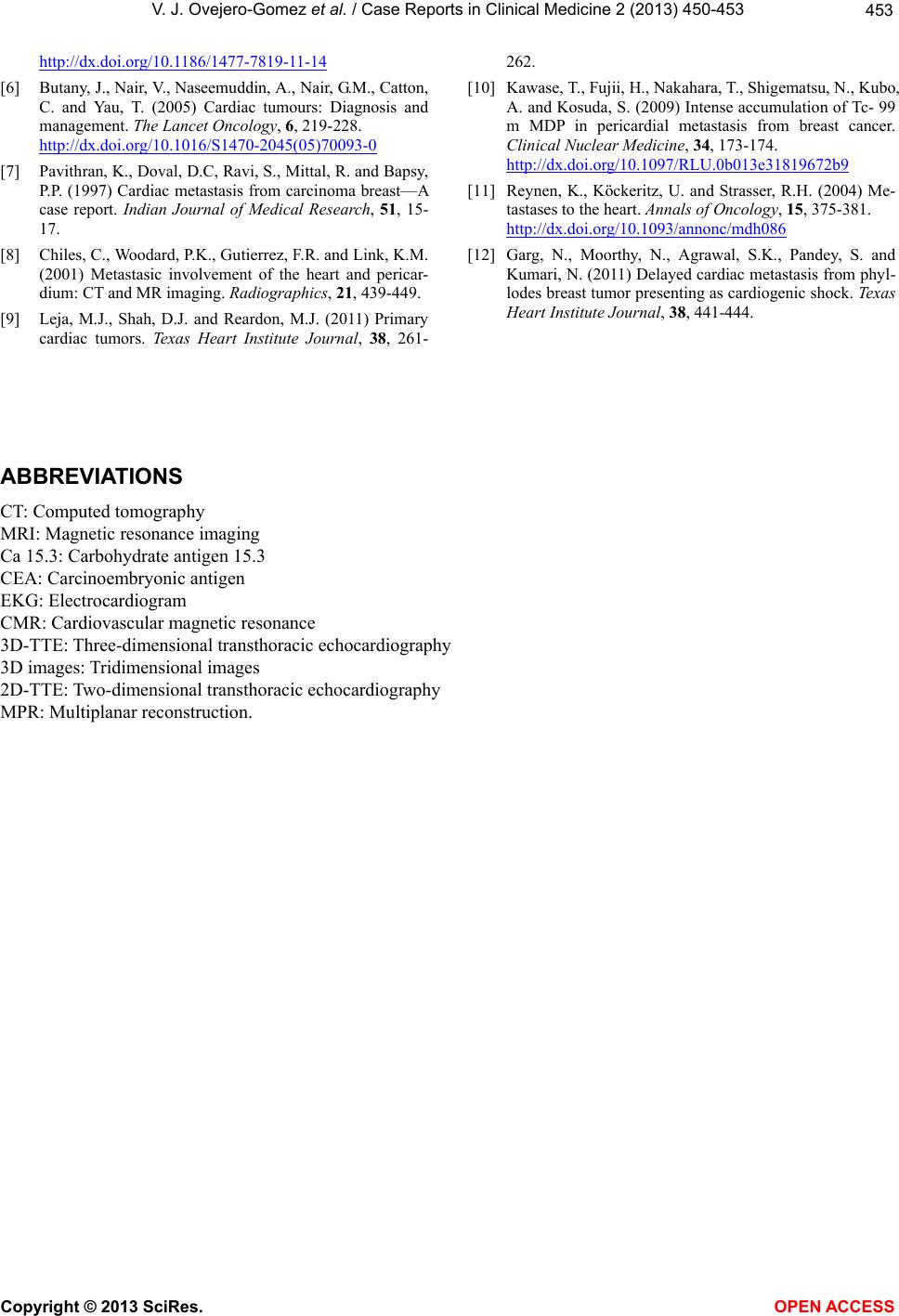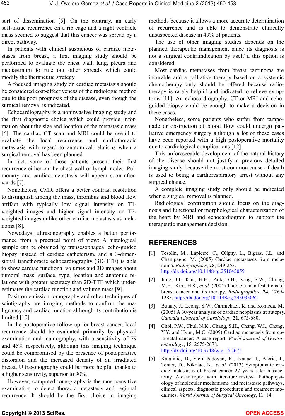
V. J. Ovejero-Gomez et al. / Case Reports in Clinical Medic ine 2 (2013) 450-453
452
sort of dissemination [5]. On the contrary, an early
soft-tissue recurrence on a rib cage and a right ventricle
mass seemed to suggest that this cancer was spread by a
direct pathway.
In patients with clinical suspicious of cardiac meta-
stases from breast, a first imaging study should be
performed to evaluate the chest wall, lung, pleura and
mediastinum to rule out other spreads which could
modify the therapeutic strategy.
A focused imaging study on cardiac metastasis should
be considered cost-effectiveness of the radiologic method
due to the poor progn osis of the disease, even thou gh the
surgical removal is indicated.
Echocardiography is a noninvasive imaging study and
the first diagnostic choice which could provide infor-
mation about th e size and locatio n of the metastasic mass
[6]. The cardiac CT scan and MRI could be useful to
evaluate the local recurrence and cardiothoracic
metastasis with regard to anatomical relations when a
surgical removal has been planned.
In fact, some of these patients present their first
recurrence either on the chest wall or lymph nodes. Pul-
monary and cardiac metastasis will appear soon after-
wards [7].
Nonetheless, CMR offers a better contrast resolution
to distinguish among the mass, thrombus and blood flow
artifact with typically low signal intensity on T1-
weighted images and higher signal intensity on T2-
weighted images unlike other cardiac metastasis as mela-
noma [8].
Nowadays, ultrasonography enables a better perfor-
mance from a practical point of view: A histological
sample can be obtained by transesophageal echo-guided
biopsy instead of cardiac catheterism, and a 3-dimen-
sional transthoracic echocardiography (3D-TTE) is able
to show cardiac functional volumes and 3D images about
tumoral mass’ surface, type, location and anatomic re-
lations with greater accuracy than 2D-TTE which under-
estimates the cardiac function and volume mass [9].
Positron emission tomography and other techniques of
scintigraphy are imaging methods to confirm the ma-
lignancy and card iac function although its co ntribution is
limited [10].
In the postoperative follow-up for breast cancer, local
recurrence should be evaluated primarily by physical
examination and mamography, with a sensitivity of 79
and 45% respectively, although this imaging technique
could be compromised by the presence of postoperative
distorsion and the increased density of an irradiated
breast. Ultrasonography could be more helpful thanks to
a higher sensitivity, superior to 90%.
However, computed tomography is the most sensitive
examination to detect thoracic metastasis and regional
recurrence. It should be the first choice in imaging
methods because it allows a more accurate determination
of recurrence and is able to demonstrate clinically
unsuspected disease in 49% of patients.
The use of other imaging studies depends on the
planned therapeutic management since its diagnosis is
not a surgical contraindication by itself if this option is
considered.
Most cardiac metastases from breast carcinoma are
incurable and a palliative therapy based on a systemic
chemotherapy only should be offered because radio-
therapy is rarely helpful and indicated to relieve symp-
toms [11]. An echocardiography, CT or MRI and echo-
guided biopsy could be enough to make a decision in
these cases.
Nonetheless, some patients who suffer from tampo-
nade or obstruction of blood flow could undergo pal-
liative emergency surgery although a lot of these cases
have been reported with a high postoperative mortality
due to cardiological complications [12].
This unforeseeable development of the natural history
of the disease should not justify a previous detailed
imaging study because the most common cause of death
is used to being a cardiorespiratory arrest without any
surgical chance.
A complete imaging study only should be indicated
when a surgical removal is planned.
Radiological contribution should focus on the diag-
nosis and functional or morphological characterization of
the heart by MRI and echocardiogram to support the
therapeutic management decision.
REFERENCES
[1] Tesolin, M., Lapierre, C., Oligny, L., Bigras, J.L. and
Champagne, M. (2005) Cardiac metastases from mela-
noma. Radiographics, 25, 249-253.
http://dx.doi.org/10.1148/rg.251045059
[2] Jung, J.I., Kim, H.H., Park, S.H., Song, S.W., Chung,
M.H., Kim, H.S., et al. (2004) Thoracic manifestations of
breast cancer and its therapy. Radiographics, 24, 1269-
1285. http://dx.doi.org/10.1148/rg.245035062
[3] Butany, J., L eong, S.W., Carmichael, K. and Komeda, M.
(2005) A 30-year analysis of cardiac neoplasms at autopsy.
Canadian Journal of Cardiology, 21, 675-680.
[4] Choi, P.W., Chul, N.K., Chang, S.H., Chan g, W.I., Chang,
Y.Y. and Hyun, M.C. (2009) Cardiac metastasis from co-
lorectal cancer: A case report. World Journal of Gastro-
enterology, 15, 2675-2678.
http://dx.doi.org/10.3748/wjg.15.2675
[5] Katalinic, D., Stern-Padovan, R., Ivanac, I., Aleric, I.,
Tentor, D., Nikolac, N., et al. (2013) Symptomatic car-
diac metastases of breast cancer 27 years after mastec-
tomy: A case report with literature review—Pathophysi-
ology of molecular mechanisms and metastasic pathways,
clinical aspects, diagnostic procedures and treatment mo-
dalities. World Journal of Surgical Oncology, 11, 14.
Copyright © 2013 SciRes. OPEN ACCESS