Y.-C. LIN ET AL.
Copyright © 2013 SciRes. OPJ
307
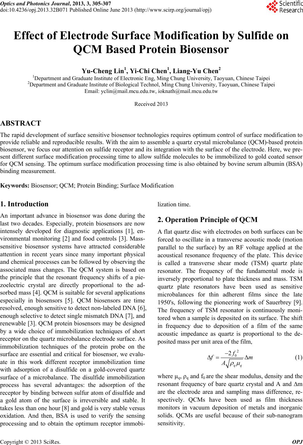
Table 1. Surface modification and protein binding effect.
chip # 1 # 2 # 3 # 4
Surface modification time (minute) 15 30 45 60
BSA binding time ( minute) 30 30 30 30
BSA frequency shift (Hz) 117.3 136.8 137.8138.0
Efficiency (%) 3.75 4.37 4.354.65
Adsorption mass (ng) 314 366 368369
4. Results and Discussion
The frequency difference after BSA injection for 4 QCM
chips with modification time of 15, 30, 45 and 60 min-
utes, respectively, is list in Table 1. Since sulfide could
bind BSA and adsorb the mass on the surface of the chip,
the resonant frequency would degrade. More BSA be
binded, larger frequency difference will be. The fre-
quency difference were measured as 117.3 Hz, 136.8 Hz,
137.8 Hz and 138.0 Hz for the 15, 30, 45 and 60 minutes,
respectively. The efficiency is defined as the frequency
difference divided with the initial deionized water reso-
nant frequency. The results are 3.75%, 4.37%, 4.35% and
4.65%, respectively. So, the optimum sulfide modifica-
tion time for BSA protein QCM biosensor is about 30
minutes.
5. Conclusions
In this study, we present different surface modification
processing time to allow sulfide molecules to be immobi-
lized to golden electrode for QCM protein sensor. The
surface modification was also tested by BSA binding
measurement. The optimum surface modification proc-
essing time is detected as 30 minutes.
REFERENCES
[1] M. Campàs and I. Katakis, “DNA Biochip Arraying, De-
tection and Amplification Strategies Trend,” Analytical
Chemistry, Vol. 23, 2004, pp. 49-62.
[2] S. Rodriguez-Mozaz, M. J. López de Alda, M.-P. Marco
and D. Barceló, “Biosensors for Environmental
Monitoring A Global Perspective,” Talanta, Vol. 65,
2005, pp. 291-297, “Title of Paper If Known,” unpub-
lished. doi:10.1016/S0039-9140(04)00381-9
[3] I. Mannelli, M. Minunni, S. Tombelli and M. Mascini
“Quartz Crystal Microbalance (QCM) Affinity Biosensor
for Genetically Modified Organism (GMOs) Detection,”
Biosensors and Bioelectronics, Vol. 18, 2003, pp.
129-140.doi:10.1016/S0956-5663(02)00166-5
[4] J. Rickert, A. Brecht and W. Gopel, “Quartz Crystal
Microbalances for Quantitative Biosensing and Character-
izing Protein Multilayers,” Biosensors and Bioelectronics,
Vol.12, pp. 567-575, Mill Valley, CA: University Science,
1989.
[5] S. Lin, C. C. Lu, H. F. Chien and S. M. Hsu. “An On-line
Quantitative Immunoassay System Based on A Quartz
Crystal Crobalance,” Journal of Immunological Methods,
Vol. 239, No.1-2, 2000, pp. 121-124.
doi:10.1016/S0022-1759(00)00184-8
[6] Y. Okahata, Y. Matsunobu, K. Ijiro, M. Mukae, A. Mu-
rakami and K. Makino, “Hybridization of Nucleic Acids
Immobilized on A Quartz Crystal Microbalance, Journal
of the American Chemical Society,Vol.114,1992,pp.
8299-8300.doi:10.1021/ja00047a056
[7] F. Höök, A. Ray, B. Nordén and B. Kasemo, “Charac-
terization of PNA and DNA Immobilization and Subse-
quent Hybridization with DNA Using Acous-
tic-Shear-Wave Attenuation Measurements,” Langmuir,
Vol. 17, 2001, pp. 8305-8312.doi:10.1021/la0107704
[8] E. Huang, M. Satjapipat, S. Han and F. F Zhou, “Surface
Structure and Coverage of An Oligonucleotide Probe Teth-
ered onto A Gold Substrate and Its Hybridization Effi-
ciency for A Polynucleotide Target,” Langmuir, Vol. 17
2001, pp. 1215-1224.doi:10.1021/la001019i
[9] G. Sauerbrey, Z. Physik, Vol. 155, 1959, p.
206.doi:10.1007/BF01337937