Keywords: Arythenoid; Hyoid Bone; Anatomic Variation; Larynx
1. Case Presentation
A 15-year-old girl with a foreign body sensation in her throat was referred for examination.
No foreign body was observed and her vocal cords showed good movement.
Following an endoscopic examination, we found a round bulge near the right arythenoid: Is this a “third arythenoid?” (Figure 1).
CT scan of the neck showed an elongated and curved right side of the hyoid bone, extending to the right arythenoid area (Figure 2 and 3).
This finding did not disturb the vocal box. Despite the bulge’s appearance as a “third arythenoid”, it actually was an anatomical variant projection of the hyoid bone into the supraglottic area.
2. Discussion
The hyoid bone is a horseshoe-shaped bone which lies between the thyroid cartilage and the mandible, consisting of the body and two pairs of cornua—the greater and the lesser cornu. The greater cornu projects posterosuperiorly as it extends laterally from the body.
It is the only bone in the human skeleton with no articulation to any other bone [1].
Over-extended bending of the greater cornu may project to the lumen of the supraglottic area as the patient presented.
Anomalies of the digastric muscle and the thyro-hyoid articulation have been described [2,3] in a single report from France [4]. The authors reported the developmental
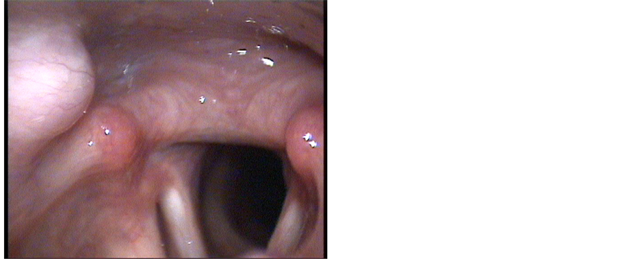
Figure 1. Endoscopic view of the larynx of the patient showing a rounded bulging near the right arythenoid, that does not move as arythenoids do during speaking but moves wile swallowing.

Figure 2. Axial CT scan of the neck shows an elongation and curve on the right side of the hyoid bone, extending medially to the laryngeal area close to the right arythenoid.

Figure 3. Reconsrtuction of axial CT scan of the neck shows an elongation and curve on the right side of the hyoid bone.
anomaly of the hyoid bone with an unusual cause of dysphagia. They described a hyoid syndrome caused by a developmental anomaly of the second branchial cleft, presenting in an adult with dysphagia.
3-D CT of the hyoid bone anomaly showed an uncurvated and elongated lesser cornu, causing persistent impingement on the lateral oropharynx wall.
This case is similar to our patient’s case, which is the second case report on the topic, to the best of our knowledge. It is the first report on greater cornu anomaly, presented as “a third arythenoid.”
Acknowledgment
Reuven Schreiber, Radiology Department, Rambam Medical Center, Haifa.
[2] H. Gray, “The Hyoid Bone,” Anatomy of the Human Body, II. Osteology, 5b. 9.
[3] A. Holiblova and L. Machalek, “A Report on Anomalies of the Digastric Muscle,” Acta Universitatis Palackianae Olomucensis Facultatis Medicae, Vol. 142, 1999, pp. 57- 59.
[4] V. Ilankovan, “An Anomaly of the Thyro-Hyoid Articulation,” The Journal of Laryngology & Otology, Vol. 101, No. 9, pp. 959-961. http://dx.doi.org/10.1017/S0022215100103068
[5] A. Boyadjian, K. Marsot-Dupuch, E. Scmitt, C. Chouard and J. Tubiana, “Developmental Anomaly of the Hyoid Bone: An Unusual Cause of Dysphagia,” Journal of Radiology, Vol. 82, No. 4, 2001, pp. 491-494.
NOTES