Effects of 4-(3-Chloro-Benzyl)-6,7-Dimethoxy-Quinazoline on Kinetics of P120-Catenin and Periplakin in Human Buccal Mucosa Squamous Carcinoma Cell Line ()
1. Introduction
Epidermal growth factor receptor (EGFR) is a receptor-type protein for tyrosine kinase [1] , and EGFR signaling controls intercellular processes including growth, differentiation or migration in epithelial cells. EGFR is colocalized with E-cadherin [2] in the cell membrane area of polarized cells [3] -[5] . Numerous studies have revealed that adhesion by E-cadherin are inhibited by EGFR activation [6] [7] , and that tyrosine phosphorylation of the E-cadherin and catenin families reduce cell-cell adhesion [8] . On the other hand, one of the earliest molecular events resulting from engagement of E-cadherin is the rapid and sustained activation of the MAPK cascade though EGFR [3] , E-cadherin-catenin complexes also induce EGFR tyrosine phosphorylation [5] . E-Cadherin is involved in Ca2+-dependent cell-cell adhesion, while the catenin family is connected to the cytoplasmic domain of cadherins via cytoplasmic α-actin, and plays an important role in intercellular adhesion. In particular, β-catenin binds to E-cadherin and links the cadherin complex to the actin cytoskeleton through α-catenin [9] . P120- catenin binds to cadherin and regulates surface levels of E-cadherin by modulating cadherin turnover [10] . As a result of the formation of the cadherin-catenin cell-cell adhesion complex, epithelial cells form layers of contiguous cells crucial to maintaining epithelial tissue structure. In cancer cells, p120-cateninis phosphorylated by the EGF-EGFR complex, and phosphorylation of p120-catenin disrupts adherens junction formation and regulation of Eand P-cadherin stability [11] . This likely requires altered kinetics of phosphorylated p120-catenin.
Periplakin is a 195-kDa protein that belongs to the plakin family of cytoskeletal linker proteins and is generally localized to the cell membrane of normal stratified squamous epithelia [12] . In addition, periplakin is localized at the cell-cell boundaries of normal epithelial squamous cells in neonatal foreskin, adult breast, oral mucosa and esophageal mucosa, thus suggesting that periplakin is associated with desmosome junctions [12] and functions as a linker of intercellular cytoskeleton and cell-cell junctions [13] [14] . In esophageal dysplasia and cancer tissues, periplakin is localized in not only cell boundaries, but also in the cytoplasm. As the expression of periplakin decreases in esophageal cancer tissue [15] , recent studies have focused on the relationship between expression of periplakin and malignancy in epithelial tissues.
4-(3-Chloro-benzyl)-6,7-dimethoxy-quinazoline,which is also known as tyrphostin 1478 (tyrphostin), is currently being used to inhibit EGFR tyrosine kinase activity and has been tested as an anti-cancer drug in vitro and in preclinical models. It has shown anti-proliferative activity in human cancer cells [16] [17] . Accordingly, inhibition of EGFR activity promotes the effects of p120-catenin and periplakin kinetics and cancer cell malignancy. However, there is little information on the role of phosphorylation of p120-catenin, or on the kinetics of periplakin and carcinogenesis in primary oral cells.
In this study, we used molecular biological and immunocytochemical techniques to investigate the kinetics of p120-catenin and periplakin in the human buccal mucosa squamous cancer cell line BICR 10 treated with tyrphostin in order to detect the relationship between p120-catenin phosphorylation, expression of periplakin and malignancy in a primary human buccal mucosa squamous cancer cell line.
2. Materials and Methods
2.1. Cell Culture
BICR 10 [18] is a primary human buccal mucosa squamous cancer cell line and was obtained from the Health Protection Agency, UK. It was cultured in Dulbecco’s Modified Eagle Medium (DMEM) supplemented with 2 mM L-glutamine, 100 μg/mL streptomycin, 100 IU/mL penicillin, 0.4 μg/mL hydrocortisone and 10% FBS in a humidified atmosphere containing 5% CO2 in air at 37˚C. In this experiment, BICR 10 was initially cultured in serum-free DMEM mixture for one day. Subsequently, BICR 10 was cultured in DMEM supplemented with 10 ng/mL human recombinant EGF (Peprotech, London, UK) and 1, 2 or 3 nM tyrphostin (Merck Millipore, Darmstadt, Germany) for one hour. Controls were treated identically without tyrphostin.
2.2. Cultured Cell Growth Assay
BICR 10 cell growth was evaluated by measuring the expression of calceinusing Cell Counting Kit-F (DOJINDO, Kumamoto, Japan). BICR 10 cells were seeded at a density of 1000 cells/well in 96-well plates, and were incubated with EGF and 3 nM tyrphostin in DMEM. After the experiment, treated cells were reacted with counting solution/PBS for one hour. Student’s paired-sample t-test was used to analyze the data, and significance was defined as p < 0.05.
2.3. Western Blot Analyses
After treated cells were washed with 1 mM Na3VO4 and 50 mM NaF included in PBS for 5 min, protein samples from stimulated BICR 10 cells in 25 cm2 tissue culture flasks were solubilized in SDS-PAGE sample buffer. Protein concentrations were determined by the BAC method, and equal amounts of protein samples were fractionated and then transferred electrophoretically onto a membrane [19] . After blocking with 1% bovine serum albumin (BSA) at 4˚C overnight, the resulting membrane was incubated with antibody against EGFR, Akt, Src or p120-catenin, as well as their phosphorylated forms, or against periplakin (Table 1), respectively, followed by labeling by the streptavidin-biotin method [20] . Expressed bands were then visualized by subsequent exposure of the membranes on X-ray film, as described previously [21] . Western blot analysis confirmed the molecular size of proteins, the specificity of antibodies, and the expression of each protein.
2.4. Immunocytochemical Analyses
BICR 10 cells were seeded at a density of 1000 cells/well in chamber-slides, and were incubated with EGF and 3 nM tyrphostin in DMEM. After the experiment, cells were fixed with acetone-ethanol for 60 min. Non-specific reactions were blocked with PBS containing 1% BSA. Treated cells were incubated with the previously described antibody (Table 1), followed by RITC-conjugated anti-immunoglobulins (DAKO A/S, Kyoto, Japan) for non-phosphorylated compounds or by FITC-conjugated anti-immunoglobulins (DAKO A/S) for phosphorylated compounds. For immunocytochemical staining for periplakin, treated cells were incubated with anti-periplakin antibody, followed by FITC-conjugated anti-immunoglobulins (DAKO A/S, Japan). All images were then observed using an all-in-one fluorescent microscopy system (BZ-9000; Keyence Japan, Osaka, Japan). Samples incubated with PBS instead of primary antibody were used as negative controls.
3. Results
3.1. Cell Culture and Growth of BICR 10 Cells
BICR 10 cells appeare dirregularly shaped in culture (Figure 1(A), arrowheads). Anti-human cytokeratinanti body (KL-1) reacted in cytoplasm of BICR 10 (Figure 1(C) and Figure 1(D)). Expression of calcein was directly correlated with tyrphostin treatment (Figure 2). Expression of calceindecreased in BICR 10 cells treated with tyrphostin.
3.2. Western Blot Profiles
Expression of periplakin and EGFR, Akt, Src and p120-catenin as their phosphorylated proteins were investigated by performing Western blot analyses, as shown in Figure 3. Although changes were not observed in the expression of EGFR and p120-catenin, the expression of Akt, Src and periplakin in BICR 10 treated with 3 nM tyrphostin tended to decrease (brackets). There was inhibition in the phosphorylation of EGFR, Akt and Src, as well as inhibition in protein kinase activity. In addition, the phosphorylation of p120-catenin was inhibited.
3.3. Immunocytochemical Observations
Immunocyto chemical staining was performed for protein localization (Figures 4-8). EGFR was localized in the

Table 1. The list of antibodies used.

Figure 1. Phase microscopy (A); staining by DAPI (B) and expression of cytokeratin (C) and (D) in BICR10. Although irregularly shaped BICR 10 cells were partially observed (A; arrowheads), pavement-shaped BICR 10 cells grew in culture. Strong reactions for KL-1 are seen in the cytoplasm of BICR 10.

Figure 2. Effects of 3 nM tyrphostin on growth of BICR10. Calcein content in BICR10 measured at O.D. 540 nm. Tyrphostin inhibits growth of BICR10.
cell membrane and was diffusely present in the cellular plasma. In addition, immunoreactions for phosphorylated EGFR were weak in BICR 10 treated with tyrphostin (Figure 4). Akt (Figure 5) and Src (Figure 6) were localized in the cellular plasma. Treatment with tyrphostin decreased immunocytoreactions against phosphorylated Akt. P120-catenin stained along the cell membrane (Figure 7, arrowheads). Weak immunocytoreactions against phosphorylated p120-catenin were observed in BICR 10 treated with tyrphostin (Figure 7, arrow). Periplakin was mainly localized along the cell membrane (Figure 8, arrowhead), and partly in the cellular plasma

Figure 3. Expression of periplakin, EGFR, Akt, Src and p120-catenin and phosphorylation of EGFR, Akt, Src and p120-catenin using Western blot analyses. In BICR10 treated with tyrphostin, expression of Akt, Src and periplakin were inhibited. Although changes were not observed in the expression of EGFR and p120-catenin, the phosphorylation of EGFR, Akt and Src and p120-catenin tended to decrease.

Figure 4. Expression of EGFR (200×). Scale bar is 50 um. Immunocytochemical reactions to EGFR or phosphorylated EGFR were diffuse. By treating with tyrphostin, BICR10 decreased the phosphorylation of EGFR. Both BICR10 cells showed no changes in expression of EGFR.
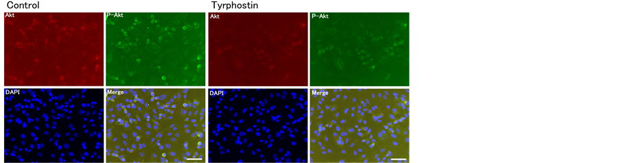
Figure 5. Expression of Akt (200×). Scale bar is 50 um. Expression and phosphorylation of Akt decreased by treatment with tyrphostin.
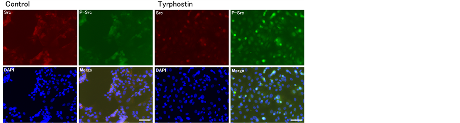
Figure 6. Expression of c-Src (200×). Scale bar is 50 um. Although the expression of c-Src decreased with tyrphostin treatment, clear changes were not observed with regard to phosphorylation of c-Src on immunocytochemical staining.

Figure 7. Expression of p120 catenin (200×). Scale bar is 50 um. Anti-p120- catenin antibody reacted along the cell membrane (arrowheads). Although both BICR10s showed no changes in expression of p120-catenin, BICR10 treated with tyrphostin decreased the phosphorylation of p120-catenin (arrow).
(Figure 8, arrow). There was a slight immunocytochemical reaction against periplakin in BICR 10 cells induced with tyrphostin.
4. Discussion
It has been revealed that activation, over expression or mutation of EGFR is related to cancer progression and poor prognosis [22] . In addition, inhibition of EGFR functions, such as autophosphorylation or signal transduction, affects cancer cell growth, infiltration and metastasis [23] -[26] , and represents an attractive method for cancer therapy, with preclinical utility being confirmed in combination with chemotherapy and radiotherapy [27] . Accordingly, it is important to establish molecular markers for therapeutic effects and prognosis in cancer.
It has been confirmed that epithelial mesenchymal transition (EMT) reflects the characteristics of cancer and
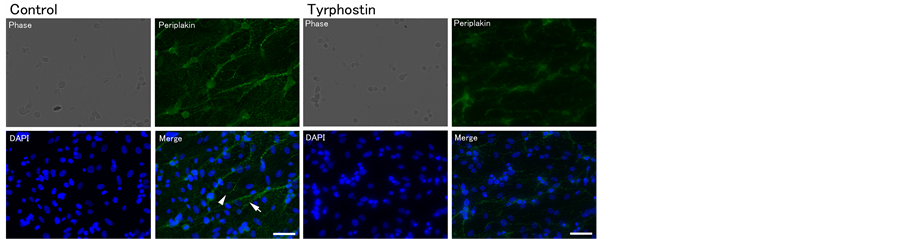
Figure 8. Expression of periplakin (200×). Scale bar is 50 um. On immunocytochemical staining, expression of periplakin showed no changes with tyrphostin treatment.
is related to cancer malignancy [28] [29] . As EMT is determined by the kinetics of adhesion molecules and skeletal proteins, it is likely that investigation into the kinetics of adhesion molecules such as p120-catenin and periplakin in cancer cells treated with tyrphostin are available to diagnosis the pharmacological effects of tyrphostin.
For these concepts, we investigated the kinetics of Akt and Src, which are known to be downstream molecules of EGFR-related signal transduction, and the kinetics of p120-catenin and periplakin, which control scancerous infiltration and metastasis, during cultivation with tyrphostin in DMEM.
Generally, inhibition of EGFR by chemical compounds other than anti EGFR anti body leads to EGFR dephosphorylation [28] . As the reaction of tyrphostin with BCIR 10was similar, BCIR 10 was characterized as being sensitive to tyrphostin. In addition, inhibition of EGFR activity by tyrphostin in BCIR 10 induced the dephosphorylation of Akt or Src downstream of EGFR transduction. These data reveal that signal transduction for survival of BCIR 10 is also EGFR-dependent. Furthermore, EGFR, Akt [30] and Src [31] are over expressed in a subset of breast cancer, which have enhanced EGFR-dependent signaling and carcinogenesis relative to other breast cancer cells that do not over express any proteins. There were no changes in the expression of EGFR, and expression of Akt or Src tended to decrease in BCIR 10 treated with 3 nM tyrphostin. These results indicate that inhibition of EGFR is not related to expression of EGFR, and that it induces low expression of Akt and Src. However, expression of periplakin, which tended to decrease after treatment with tyrphostin, may affect Akt and Src signaling.
Adherence junction molecules and related glycoproteins are involved in not only cell adhesion, and but also signal transduction [21] [32] . These proteins are regulated by receptor tyrosine kinases [11] [32] , and p120-cateninis strongly expressed by general potentially malignant oral cells and is phosphorylated at the 228th tyrosine [33] . As the phosphorylation of p120-catenin was inhibited by tyrphostin on Western blotting analysis, it may be useful as a biomarker of treatment effects on EGFR inhibitor.
5. Conclusion
In conclusion, the decrease in phosphorylation of EGFR and p120-catenin by tyrphostin, following the decrease in Src or Akt phosphorylation, may inhibit expression of several growth factors associated with the proliferation and migration of cancer cells.
Acknowledgements
This study was performed using the Morphological Research Facilities, Low Temperature Facilities, Photograph-Processing Facilities, Analytical Instrument Facilities and Dental Bioscience Facilities I of the Institute of Dental Research, Osaka Dental University.